

Key Points
The sacroiliac joints join the pelvis to the spine
A blow to the hind end can result in dislocation of one or both sacroiliac joint
This can result in severe lameness or nonweight-bearing lameness of the hind limb(s)
Sciatic nerve damage can occur in cases that sustain a sacroiliac joint dislocation
Other concurrent injuries that must be ruled out include rupture of the bladder, rupture of the lung, and internal hemorrhage from trauma to the spleen or liver
Initial visit- downloadable forms/information for clients:
History sheet - Client initial consultation history
Client education handout - pelvic fractures
Discharge instructions for postop care - dog
Discharge instructions for postop care - cat
2-week recheck visit - downloadable forms/information for clients:
History form - 2 week postop (Telemedicine evaluation) - dog and cat
Postop care instructions 2-week fracture repair recheck - cat
Postop care instructions 2-week fracture repair recheck -dog
8-week recheck visit - downloadable forms/information for clients:
History form 8-week fracture recheck (in hospital) - dog and cat
Postop care instructions 8 week fracture recheck - dog
Downloadable forms for referring veterinarians:
Anatomy
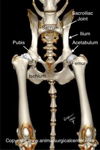
The pelvis consists of two symmetrical sets of bones that are fused together to form a solid bone. The pelvis is attached to the lower part of the spine, called the sacrum, by a a left and right sacroiliac joints. Unlike the hip, knee, elbow and other joints, the sacroiliac joints have limited movement. The sacrum consists of three vertebral bones that are fused together
The sciatic nerve runs immediately below the sacroiliac joint, thus this nerve is susceptible to damage
Signs
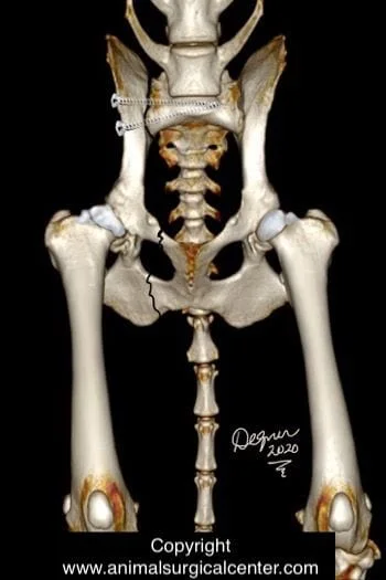
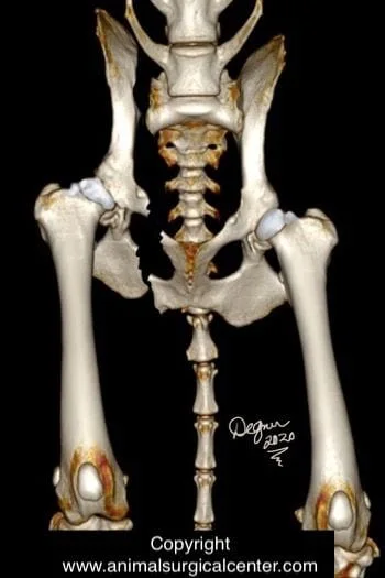
Sacroiliac luxation (luxation = dislocation in layman’s terms) is a traumatic condition in which the pelvis has been torn off the spine; concurrently the pelvis is typically also broken in one or two other locations (typically pubis and ischium). Bilateral sacroiliac luxation may occur with no additional fractures of the pelvis. Sacroiliac luxation usually is caused by a very traumatic blow to the hind end of an animal (i.e. hit by a motor vehicle). Varying degrees of lameness on the side of the luxation. Pain is noted when the sacroiliac joint is palpated. The pelvis may feel crunchy (crepitant) when a force is applied to the front of the pelvis.
If the sciatic nerve was also injured, no or decreased sensation to the outside toe of the affected hindlimb will be noted; complete evaluation of the sciatic nerve may be difficult due to bruising of muscles and the nerve itself.
Diagnosis
Prior to surgery a complete blood count and chemistry profile will be performed. Radiographs (x-rays) of the chest will be completed to rule out trauma to the lungs or ribs. Radiographs or ultrasound of the abdomen to rule out internal organ damage and internal bleeding. Your pet's surgeon may also consider performing a whole body CT scan to more thoroughly examine the pet.
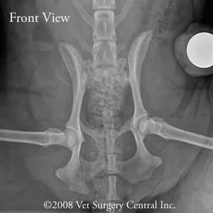
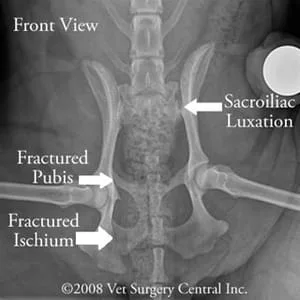
Radiographs of the pelvis; the unfractured pelvis should fit within an imaginary box and if it does not, there is a fracture +/- dislocation the sacroiliac joint a part of the pelvis will be shifted outside this box. In this case, take note of the sacroiliac luxation and fractures of the pubis and ischium of the pelvis.
Treatment
1. Conservative treatment is acceptable if only one side of the pelvis has been damaged, and minimal displacement of the sacroiliac joint is present. These patients will be painful for a much longer time versus having the luxation surgically repaired.
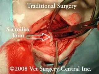
2. Traditional surgery involves making an incision along the side of the pelvis and peeling the muscles off the bones to expose the dislocated joint (photo right). Screws are used to secure the dislocated joint in place.
3. Minimally invasive surgery involves manually reducing the joint back into place from the outside, making a small incision (about 1 to 1.5 cm) over the side of the pelvis in the location of the sacroiliac joint and securing the sacroiliac joint in place with screws with the aide of intra-operative fluoroscopy or digital radiography.
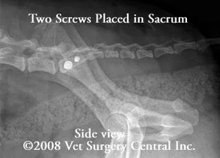
How minimally invasive sacroiliac laxation is performed: The patient is placed on the x-ray table with the affected pelvis up. Digital radiographs are made (or fluoroscopy used) to assist the surgeon in the placement of the screws. The ilium is manually pushed back into place and a small pin is inserted. Next, a hole is drilled over sacral body and the position of the drill bit is radiographed. If the positioning is good, the drill bit is advanced deeply into the sacrum. The drill bit is removed and a screw (premeasured screw length from the radiographs) is seated into the bones to hold the sacroiliac joint in place. Another screw is placed just behind the first screw using the same method.
The benefits of the minimally invasive approach is that a very small incision is used and there is minimal dissection of the soft tissues, resulting in less pain. In addition, the surgeon can very accurately place the screws into the body of the sacrum, thereby preventing damage to the nerves that are located in the spinal canal of the sacrum. The length of time that it takes to perform the minimally invasive versus traditional surgery is similar.
The radiographs below were taken during surgery.


Home care
Limit activity until the fractures have healed. Provide a soft bed to prevent bed sores. Turn your pet from side to side, if your pet is very weak and cannot do this. Use a towel as a sling to take your pet outside for elimination purposes. Check the incision for infection.
Potential complications
Complications of the condition of sacroiliac luxation or surgery to repair the luxation may include sciatic nerve damage, nonhealing of the fractures, breakage of the screws, infection, and anesthetic death. Pregnancy should be avoided as natural delivery of the puppies may not be possible due to scar tissue or callus formation in the pelvic canal (C-section is also an option if breeding is a must). Chronic constipation may occur if a lot of callus or scar tissue develops in the pelvic canal. Entrapment of the urethra (tube from the bladder for urination) may occur by entrapment of this tube by fracture fragments.
Prognosis
Most patients heal well following surgical repair. It may take about 2 months for full recovery. Patients that have sciatic nerve injury frequently will regain normal or near normal function; occasionally the function of the nerve does not come back and the pet does not have any useful function of the limb.
