

Key Points
- Elbow fractures occur most commonly in young dogs
- Surgery should be done soon after the fracture has occured
- Accurate surgical reduction of the fracture results in the best outcome
- Some arthritis may develop in the elbow joint in the future, but is minimized by having surgery done

Downloadable forms/information for clients for initial visit:
Client initial consultation history sheet
Elbow fracture client education handout
Discharge instructions for elbow fracture postop care
Downloadable forms/information for clients for 2-week recheck visits:
2-week fracture recheck (Telemed)
Downloadable forms/information for clients for 5 to 8-week recheck visits:
5 to 8-week fracture recheck (in hospital)
5 to 8-week fracture discharge instructions
Downloadable forms for referring veterinarians:

Anatomy
The elbow joint is made of three bones that are joined together by ligaments, tendons and muscles. These bones include the humerus, the radius and the ulna. The radius bone bears 80% of the weight exerted on the forelimb during weight-bearing, while the ulna bears 20% of the weight. The head of the radius articulates with the lateral or outer one-half to two-thirds of the humerus bone. The ulna bone articulates with the inner portion of the humerus bone.
Why do elbow fractures occur?
An impacting force such as jumping off a deck, chair or out of a pet owner's arms may cause this type of fracture. The most common type of elbow fracture is a lateral condylar fracture of the lower part of the humerus bone. Because forces are driven up the axis of the radius bone, the radial head will impact the lateral condyle of the humerus bone and shear the lateral condyle off the humerus bone. Young dogs, approximately 4 to 5 months of age, are most susceptible to this type of fracture, because the bone is soft and the medial and lateral condyles have not fused together. In mature dogs, this fracture also is seen and the predisposing factor usually is due to incomplete ossification of the humeral condyles (IOHC). This condition is likely a genetic disease.
Incomplete ossification of the humeral condyles
Early on in life, the bones of a fetus are made of cartilage. As the puppy matures, the bones gradually change from a cartilage primordia by a process of ossification (or mineralization). The bottom of the humerus has two condyles, the medial and the lateral condyle which have separate ossification centers. Thus, as a puppy grows, the ossification of the two condyles progress toward each other and finally fuse together into a solid bone. In some dogs, due to a genetic defect, the medial and lateral condyles never fuse together completely and a fine line of cartilage separates the two. This is a weak link between the two condyles, which makes this region vulnerable to fracture. The radius bone, being the main weight-bearing bone drives its forces upon the lateral condyle. Because of this, the lateral condyle may be fractured off the humerus. Below are illustrations demonstrating the progression of the fracture from just a fissure in the distal humerus between the medial and lateral condyles, to propagation of the fracture in the lateral epicondylar crest, then finally a complete lateral condylar fracture.
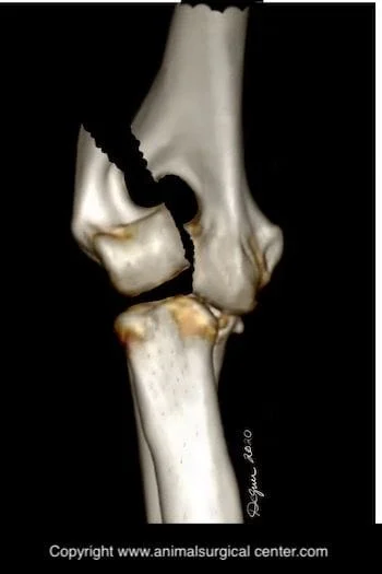
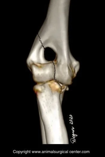
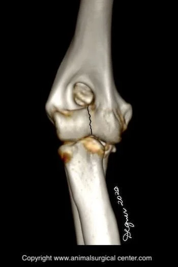
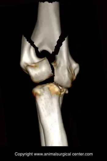
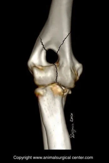
Types of distal humeral fractures
There are three main types of distal humeral fractures that we seen in our practice. From most common to least common, the lateral condylar fracture, "Y" supracondylar fracture, medial condylar fracture. The "Y" supracondylar fracture splits both medial and lateral condyles off the distal humerus and this is a very challenging fracture to repair. The lateral condylar fracture is shown above and the "Y" fracture is shown below.
Clinical signs
Fracture of the humerus bone results in extreme initial pain and the pet may cry out during the injury. These patients typically do not bear any weight on the fractured limb. Movement of the limb typically will result in pain and the elbow may feel unstable or you may feel crackling of broken bones.

Diagnosis
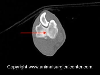
The diagnosis of an elbow fracture is quite obvious on x-rays of the limb. CT scan may be recommended by your pet's surgeon to see if the opposite humerus has IHOC. If this is the case, then fracture of this humerus is also possible in the future. These two CT scan images show a fine crack between the medial and lateral humeral condyles, making this dog prone to a humeral fracture. If your pet sustained major trauma (hit by car) that caused the fracture, chest x-rays and potentially a body CT scan will be recommended by one of our surgeons. Blood work is done prior to surgery to ensure that the internal organs are functioning well and your pet is not anemic.
Preparation for surgery
The pet should be fasted prior to surgery, as instructed by the surgical team. Water is usually permitted up to the time of admission to the hospital. The surgical team should be informed of any medications that your pet is currently receiving. Just prior to surgery, your pet will receive a sedative, have an intravenous catheter placed for the administration of intravenous fluids and intravenous medications, be induced under general anesthesia with medication(s), and have a breathing tube (endotracheal tube) placed to allow delivery of oxygen and gaseous anesthesia. The surgical site will be clipped and cleansed with an anti-septic solution in preparation for surgery. While under general anesthesia, the pet’s breathing will be assisted with a ventilator and vital parameters such as heart rate, respiratory rate, core body temperature, blood pressure, oxygenation of the blood (pulse oximetry), exhaled carbon dioxide (capnography), and heart rhythm (EKG) will be monitored to ensure the pet’s well being. Pain will be controlled both during and after surgery with analgesics (pain-controlling medication). Please note that each surgical and anesthesia team may elect to chose a different, but effective analgesia protocol.
Surgery
If the fracture is not repaired soon after injury, developing scar tissue can make accurate surgical reduction of the fracture much more difficult. It is vital that the surface of the joint is lined up well to minimize arthritic changes. If the elbow fracture is not repaired, the dog will develop debilitating arthritis.
During the surgery, the surgeon will make an incision on the lateral or outer side of the elbow if a lateral condylar fracture is present. If a "Y" fracture is present, either a large incision on the lateral side of the elbow combined with transection of the olecranon or an incision on both the medial and lateral sides of the limb are made.
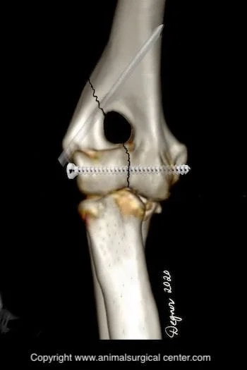
For a lateral condylar fracture, the lateral condyle is reduced in place and a transcondylar screw is placed followed by a pin which extends from the lateral epicondyle to the shaft of the humerus.
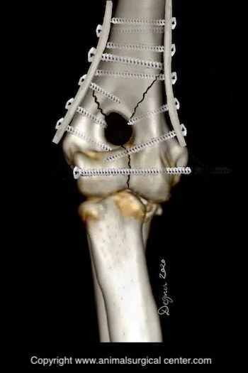
For a "Y" fracture fracture, the medial and lateral condyles are reduced together and a transcondylar screw is placed. After that, two metal plates are placed on each side of the bone and secured with a series of screws. Instead of two metal plates, the surgeon may use a series of pins or plate and pin.
Potential complications of surgery and or the condition of a fractured elbow may include anesthetic death, infection, seroma, breakage of the plates/pins or screw(s), failure of the bones to heal, reduced range of motion, arthritis, and nerve damage.
Aftercare
Prescribed medication to prevent infection and control pain are to be administered to your pet after surgery. Exercise should be restricted until the bone is deemed to be healed, based on x-ray images. Passive range of motion exercises are important to achieve normal range of motion of the elbow joint. Your pet will also benefit from therapy from a professional rehabilitation therapist.
Success
After surgery, about 80% of the patients do very well after surgery. It is imperative that the discharge instructions are closely followed to reduce postoperative complications that may have long-term devastating effects on the outcome.
