

Key Points
The lower (distal) part of the femur bone has a growth plate which is susceptible to fracturing in puppies and kittens
Surgery is needed to correct this type of fracture
Prognosis with surgical correction is favorable
Initial visit- downloadable forms/information for clients:
History sheet - Client initial consultation history
Distal Femoral Physeal fracture client education handout
Discharge instructions for postop care - dog
Discharge instructions for postop care - cat
2-week recheck visit - downloadable forms/information for clients:
History form - 2 week postop (Telemedicine evaluation)
Postop care instructions 2-week Fracture repair recheck - dog
8-week recheck visit - downloadable forms/information for clients:
History form 8-week fracture recheck (in hospital)
Postop care instructions 8 week fracture recheck
Downloadable forms for referring veterinarians:

Basic facts
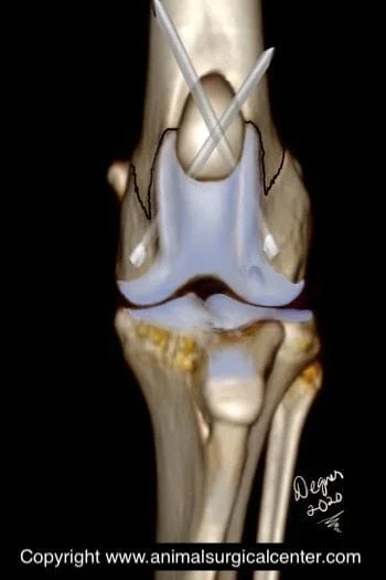
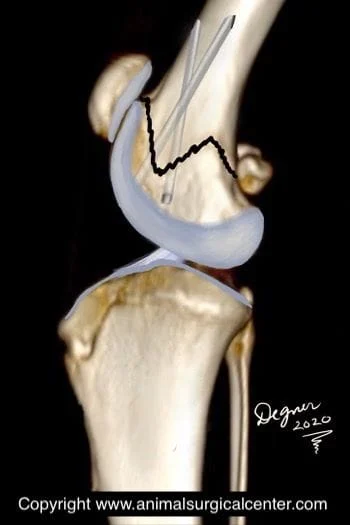
A fracture is synonymous with a broken bone. Puppies and kittens have growth plates at the end of the femur bone from which the bone grows. Growth plates are susceptible to developing fractures in immature animals. One such growth plate located at the bottom of the femur bone called the distal femoral physis.
Signs and diagnosis
This type of fracture usually occurs in dogs and cats that are less than one year of age. The most common cause of a femoral fracture is trauma such as being struck by a motorized vehicle or taking a fall. Affected pets will bear minimal to no weight on the fractured limb. The lower part of the thigh, near the knee will be swollen, painful to touch and may be crepitant (crunchy) when the knee is flexed and extended. The diagnosis of this type of fracture is made by x-raying the limb.


Preparation for surgery
The pet should be fasted prior to surgery, as instructed by the surgical team. Water is usually permitted up to the time of admission to the hospital. The surgical team should be informed of any medications that your pet is currently receiving. Just prior to surgery, your pet will receive a sedative, have an intravenous catheter placed for the administration of intravenous fluids and intravenous medications, be induced under general anesthesia with medication(s), and have a breathing tube (endotracheal tube) placed to allow delivery of oxygen and gaseous anesthesia. The surgical site will be clipped and cleansed with an anti-septic solution in preparation for surgery. While under general anesthesia, the pet’s breathing will be assisted with a ventilator and vital parameters such as heart rate, respiratory rate, core body temperature, blood pressure, oxygenation of the blood (pulse oximetry), exhaled carbon dioxide (capnography), and heart rhythm (EKG) will be monitored to ensure the pet’s well being. Pain will be controlled both during and after surgery with analgesics (pain-controlling medication). Please note that each surgical and anesthesia team may elect to chose a different, but effective analgesia protocol.


Surgery
Distal femoral physeal fractures always require surgery for a successful outcome. Without surgery the pet usually will develop a stiff limb that has poor function. This type of fracture is also not amenable to a splint or cast. Surgery should be done as soon as possible, so that the broken end of the bone does not get worn down, thus preventing the fracture from fitting together properly. In addition, chronic fractures may be very difficult to put back together, as the tissues around the bones become permanently contracted. Surgical repair of a distal femoral physeal fracture requires an incision located on the outer side of the lower thigh and knee. The bones are realigned and two pins are inserted to keep the fracture in place.
Aftercare
After surgery, you can continue to give your pet a prescribed pain reliever to minimize discomfort. It’s also extremely important to limit your pet’s activity and exercise level during this post-operative period. Rehabilitation exercises can be done at your home or if you choose, by professionally trained therapists at an animal rehabilitation center. Rehabilitation therapy should be continued until your dog is bearing weight well on the operated limb (typically 2 – 4 weeks after surgery). Detailed instructions will be given to you after the surgery. The surgeon will monitor the healing process with two follow-up exams. The first is scheduled at two weeks after the surgery and the second is at five to eight weeks after the surgery; during the second visit the repaired bone will be x-rayed. By 8 weeks after surgery, most dogs and cats are fully weight-bearing on the operated limb, although exercise should be limited during the first three months after the procedure.
Prognosis
Primary repair of the fracture has been reported to be successful in 93% of the cases, whereas, no surgery will result in persistent lameness and a poorly functioning limb. Uncommon complications include infection, breakage of the pins, migration of the pins (requiring removal or replacement of the pins) and collapse of the fracture site.
