

Initial visit- downloadable forms/information for clients:
History sheet - Client initial consultation history
Capital Physeal fracture client education handout
Discharge instructions for postop care
2-week recheck visit - downloadable forms/information for clients:
Postop care instructions 2 week -FHO recheck (Telemed) - dog
Postop care instructions 2-week FHO recheck (Telemed) - cat
Postop care instructions 2-week Fracture repair recheck - dog
8-week recheck visit - downloadable forms/information for clients:
History form 8-week fracture recheck (in hospital)
8-week fracture discharge instructions
Downloadable forms for referring veterinarians:
Key Points
Fractures of the hip occur in immature cats and dogs
Surgery is the best option to repair hip fractures
The prognosis is fair to good with this type of surgery
Anatomy
Puppies have growth plates at the ends of the femur bones from which the bone grows. Growth plates are susceptible to developing fractures in immature animals. One such growth plate is called the capital physis or growth plate of the head of the femur bone. This head forms the so-called ball of the hip joint.
Cause of fracture
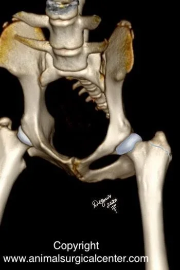
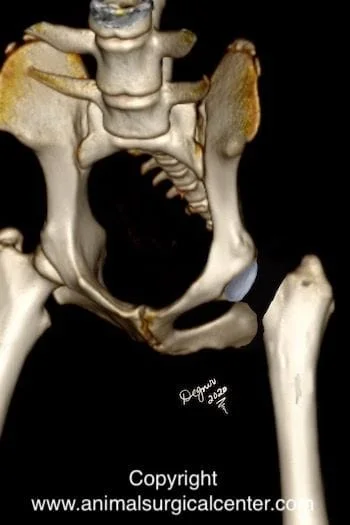
A fracture is synonymous with a broken bone. When a fracture occurs through this growth plate the cap of the head of the femur slips off, hence this fracture is referred to as a “slipped cap”. The growth plate of the head of the femur is located at the junction of the cartilage and the bone (see illustration right ). This type of fracture only occurs in dogs that are less than one year of age and cats that are just under 2 years of age. The most common cause of a femoral fracture is trauma such as being struck by a motorized vehicle or taking a fall.
Signs
Sudden onset of severe to nonweight-bearing lameness is common in patients sustaining a slipped capital physeal fracture. Palpation of the hip will elicit pain and a grating or clicking sensation may be felt when moving the hip joint.
Diagnosis
The diagnosis of a slipped capital physeal fracture requires x-rays; typical x-ray views include the hip extended, frog-leg, and lateral views. A misdiagnosis of a slipped cap can be made on the typical hip extended x-ray of the pelvis, as the hip sometimes may look fairly normal on this view. Therefore, a frog-leg view (limbs splayed apart) should be used to assess the hip joint. Other testing that may be recommended prior to anesthesia includes a complete blood count and chemistry profile to ensure that the pet is healthy. If the fracture was due to trauma such as being hit by a motor vehicle, chest x-rays should be made to rule out lung and chest trauma.
Preparation for surgery
The pet should be fasted prior to surgery, as instructed by the surgical team. Water is usually permitted up to the time of admission to the hospital. The surgical team should be informed of any medications that your pet is currently receiving. Just prior to surgery, your pet will receive a sedative, have an intravenous catheter placed for the administration of intravenous fluids and intravenous medications, be induced under general anesthesia with medication(s), and have a breathing tube (endotracheal tube) placed to allow delivery of oxygen and gaseous anesthesia. The surgical site will be clipped and cleansed with an anti-septic solution in preparation for surgery. While under general anesthesia, the pet’s breathing will be assisted with a ventilator and vital parameters such as heart rate, respiratory rate, core body temperature, blood pressure, oxygenation of the blood (pulse oximetry), exhaled carbon dioxide (capnography), and heart rhythm (EKG) will be monitored to ensure the pet’s well being. Pain will be controlled both during and after surgery with analgesics (pain-controlling medication). Please note that each surgical and anesthesia team may elect to chose a different, but effective analgesia protocol.
Surgery
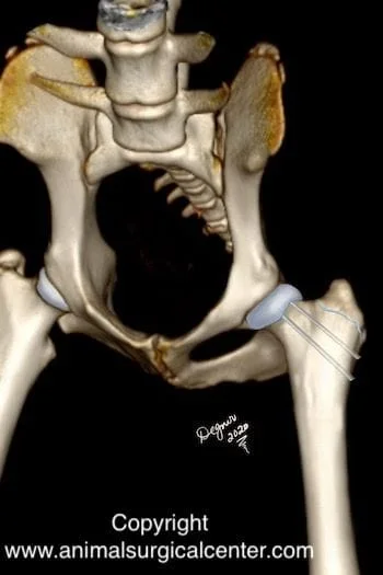
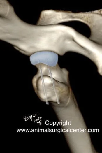
Slipped cap fractures always require surgery for a successful outcome. The surgery should be done as soon as possible so that broken end of the bone does not get worn down, thus preventing the fracture from fitting together properly. In medium to large dogs, primary repair of the fracture is always recommended. Generally two or three pins are inserted in the bone to stabilize the fracture.
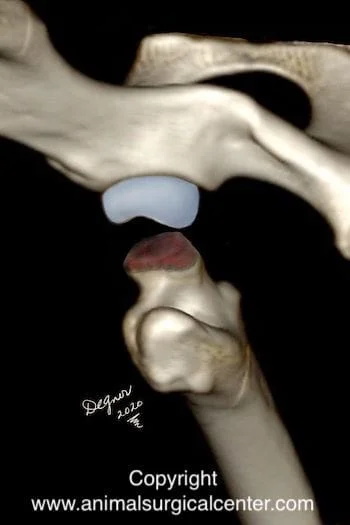
In small dogs and cats, femoral head and neck osteotomy can be performed to treat a slipped capital physical fracture and the results are usually good. In this procedure, the piece of broken bone (femoral cap) is removed and the remaining portion of the femoral neck is excised (see illustration right.
Aftercare
After surgery, you can continue to give your pet a prescribed pain reliever to minimize discomfort. It’s also extremely important to limit your dog’s activity and exercise level during this post-operative period. A sling will be placed on your companion’s hind limb to prevent weight-bearing during the first 10-14 days after surgery. Rehabilitation exercises can be done at your home or if you choose, by professionally trained therapists at an animal rehabilitation center. Rehabilitation therapy should be continued until your dog is bearing weight well on the operated limb (typically 2 – 4 weeks after surgery). Detailed instructions will be given to you after the surgery. The surgeon will monitor the healing process with two follow-up exams. The first is scheduled at two weeks after the surgery and the second is at eight weeks after the surgery. By 8 to 10 weeks after surgery, most dogs are fully weight-bearing on the operated limb, although exercise should be limited during the first three months after the procedure.
Prognosis
Surgery usually gives an acceptable outcome; however, it is possible that the dog may not be a good athlete (such as a hunting dog). Dogs that are 4 months and younger tend to develop much more arthritis than older dogs with the same fracture. Uncommon complications include infection, breakage of the pins, and sciatic nerve damage. In general most small dogs and cats that receive a femoral head and neck excision surgery regain very good function on the limb. Primary repair of the fracture has been reported to be successful in 80% of the cases, whereas, no surgery will result in persistent lameness and hip pain.
References
- Gilson KL, vanEe RT, Pechman RD femoral capital physeal fractures in dogs: 34 cases. J Am Vet Med Assoc. 1991, 198(5):886-90.
- DeCamp CE, Probst CW and Thomas MW. Internal fixation of femoral capital physieal injuries in dogs: 40 cases (1979 to 1987). J Am Vet Med Assoc 1989;12 :194(12)1750-4.
- McNicholas WT, Wilkens BE, Blevins WE, et al. Spontaneous femoral capital physeal fractures in adult cats: 26 cases (1996–2001). J Am Vet Med Assoc 2002;221:1731–6.
- Fischer HR, Norton J, Kobluk CN, et al. Surgical reduction and stabilization for repair of femoral capital physeal fractures in cats: 13 cases (1998–2002). J Am Vet Med Assoc 2004;224:1478–82.
