

Key Points
- Because the tibia bone is just beneath the skin on the inner side of the limb, there is increased risk for a sharp bony fragment to penetrate through the skin; this can be prevented by supporting the limb with a splint before the surgery is performed
- Tibial fractures are usually repaired using a plate and screws, but other forms of repair are also possible
- These type of fractures do not respond well to casting
Initial visit- downloadable forms/information for clients:
History sheet - Client initial consultation history
Client education handout - tibial fractures
Discharge instructions for postop care - dog
Discharge instructions for postop care - cat
2-week recheck visit - downloadable forms/information for clients:
History form - 2 week postop (Telemedicine evaluation) - dog and cat
Postop care instructions 2-week fracture repair recheck - cat
Postop care instructions 2-week fracture repair recheck - dog
8-week recheck visit - downloadable forms/information for clients:
History form 8-week fracture recheck (in hospital) - dog and cat
Postop care instructions 8 week fracture recheck - dog
Postop care instructions 8 week fracture recheck - cat
Downloadable forms for referring veterinarians:
Anatomy
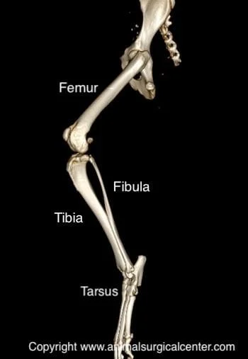
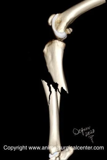
A fracture is synonymous with a broken bone. The bones between the stifle (knee) and tibiotarsal (ankle) joints are called the tibia and fibula bones (see illustration right). The top of the tibia bone has a plateau and tibial crest. The bottom of the tibia bone articulates with the talus bone. The tibia bone is well muscled which provides a good blood supply to the bone. The hind limb is innervated by the sciatic and femoral nerves. The sciatic nerve divides into the tibial nerve on the back side of the tibia and the peroneal nerve which runs along the outer and proximal side of the tibia bone and may be injured by a sharp broken bone. The femoral nerve has sensory branches that extend on the inner side of the tibia called the saphenous nerves. Puppies have growth plates at the ends of the tibia bones from which the bone grows. Growth plates are susceptible to developing fractures in immature animals.
Causes of fractures
The most common cause of a tibial fracture is trauma such as being struck by a motorized vehicle. Gunshot injuries not only will fracture the bone, but also result in a dirty open wound. This could potentially result in infection and delayed healing of the bone. If the fractured bone is sharp it may penetrate through the skin and result in infection of the bone. If the pet sustains a fracture without any known trauma, there may be an underlying disease that has weakened the bone such as a nutritional deficiency. Foods that have too much phosphorus and too little calcium or too much vitamin A will make the bones weak. Some animals have an inherited collagen defect that weakens the bones, resulting in bone fractures with minimal trauma. Bone cancer also can weaken the bone and predispose the pet to a spontaneous fracture.
Preparation for surgery
Your pet should be fasted prior to surgery, as instructed by the surgical team. Water is usually permitted up to the time of admission to our hospital. You should inform the surgical team of any medications that your pet is currently receiving and of any pertinent medical alerts (allergies, seizures, drug insensitivities, etc). Just prior to surgery, your pet will receive a sedative, have an intravenous catheter placed for the administration of intravenous fluids and intravenous medications, be induced under general anesthesia with medication(s), and have a breathing tube (endotracheal tube) placed to allow delivery of oxygen and gaseous anesthesia. The surgical site will be clipped and cleansed with an anti-septic solution in preparation for surgery. While under general anesthesia, your pet’s breathing will be assisted with a ventilator and vital parameters such as heart rate, respiratory rate, core body temperature, blood pressure, oxygenation of the blood (pulse oximetry), exhaled/inhaled carbon dioxide (capnography), and heart rhythm (EKG) will be monitored to ensure the pet’s well being. Pain will be controlled both during and after surgery with analgesics (pain-controlling medication). Please note that each surgical and anesthesia team may elect to choose a different, but effective analgesia protocol, pending the needs of the patient.
Surgery
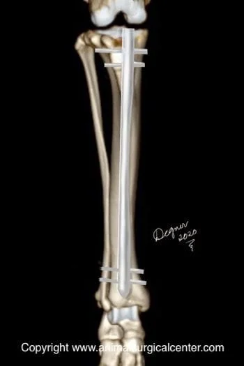
Two-piece fractures of the tibia bone are commonly stabilized with a bone plate and multiple screws (see below). Tibial fractures that are candidates for plating are those that can be reconstructed and have bone fragments that can share the weight load (i. e. plate is not the only supporting structure). If these features cannot be achieved, there is increased risk for the plate to break in the postop period, especially in a very active, rambunctious pet. The addition of a pin down the marrow cavity, in the form of a pin-plate repair, will help take stress off the metal plate.
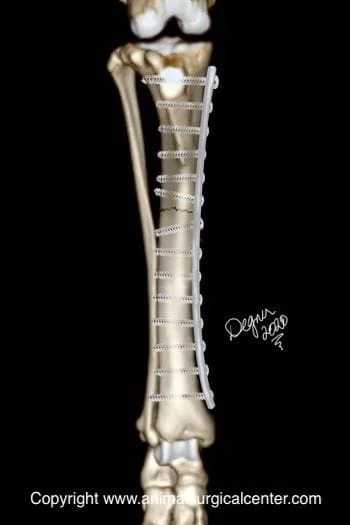
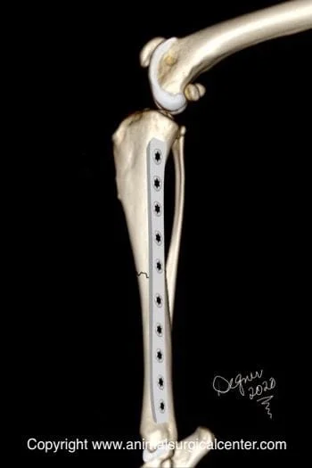
The tibia bone is very amenable to minimally invasive locking bone plating or interlocking nailing with an access incision at the top and bottom of the bone.
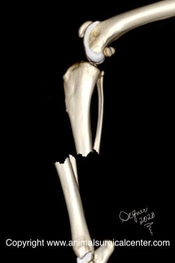
If the bone is broken in multiple pieces, called a comminuted fracture, an interlocking nail is typically the fixation apparatus of choice (see below).

Both of these treatment options provide the least aftercare for the client and a successful outcome in most cases.
If the bone fracture is associated with an open skin wound and contaminated, an external skeletal fixator may be the treatment of choice.
Tibia bone fractures can be casted or successfully splinted, however, common complications with splinting or casting include failure of the bones to heal in proper alignment and failure of the bones to heal together (called a nonunion). A maligned tibial fracture commonly result in external rotation (seal flipper) of the paw which may stress the joints of the limb.
Home care
After surgery, you can continue to give your pet a prescribed pain reliever to minimize discomfort. It’s also extremely important to limit your dog’s activity and exercise level during this post-operative period. Rehabilitation exercises can be done at your home or if you choose, by professionally trained therapists at an animal rehabilitation center. Rehabilitation therapy should be continued until your dog is bearing weight well on the operated limb (typically 2 weeks after surgery). Detailed instructions will be given to you after the surgery. The surgeon will monitor the healing process with two follow-up exams. The first is scheduled at two weeks after the surgery and the second is at eight weeks after the surgery. By 8 to 10 weeks after surgery, most dogs are fully weight bearing on the operated limb, although exercise should be limited during the first three months after the procedure.
Success
In our experience at ASCM, most fractures treated with internal reduction and fixation heal uneventfully.
