

Key Points
The dynamic tibial plateau leveling procedure (DTPLP), can be used to treat cranial cruciate ligament rupture in growing immature dogs
The timing of the surgery is critical, and is specific to the breed of dog
This technique involves placing a screw in the cranial aspect of the tibial plateau to arrest growth in this region
As the dog grows the top of the tibia levels, in essence the final result is the same as the TPLO, but with a much less aggressive surgery
Downloadable forms/information for clients for initial visit:
Client initial consultation history sheet
Discharge instructions for TPLP postop care
Downloadable forms/information for clients for recheck visits:
2 month TPLP recheck (in hospital at ASCM)
Downloadable forms for referring veterinarians:
Introduction
The stifle joint of the dog is equivalent to a human’s knee. The cranial cruciate ligament is located inside the joint and is responsible for maintaining a stable joint. One of the important functions of the ligament is to prevent backward sliding of the femur on the top of the tibia bone (called drawer motion). Cranial cruciate ligament rupture is the most common orthopedic condition that we treat at ASCM. This problem afflicts all ages and breeds of dogs. Cruciate ligament rupture is usually a progressive process and not due to a single traumatic injury. Most dogs have predisposing factors such as age-related ligament degeneration, pre-existing inflammation, anatomical abnormalities, and excessive slope of the top of the tibia bone, which weaken the ligament. Rupture of the cruciate ligament in both knees is common. In fact, 54% of dogs will rupture the cranial cruciate ligament of the opposite stifle within a 3 year period of time.
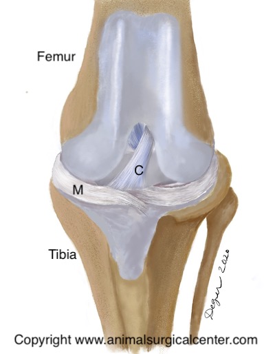
This front view of the canine stifle joint illustrates the cranial cruciate ligament (labeled C) and the medial meniscus (labeled M), which is a cartilage pad located within the stifle joint. This pad is commonly torn in dogs with cruciate ligament rupture.
Wagon Model Used to Explain Instability of the Knee Joint
The tibial plateau of a dog’s stifle is sloped. Understanding the importance of the tibial slope when the cranial cruciate ligament is torn is somewhat difficult. We therefore present a model of a wagon on a hill, which is tied to a post with a rope. The slope of the hill represents the tibial plateau, the wagon represents the femur bone, and the rope represents the cranial cruciate ligament. If the rope is torn, the wagon will roll down the hill (see fig below). Likewise, when cranial cruciate ligament is torn the femur bone slides down the slope of the tibial plateau. When surface that the wagon is placed on is leveled and weight is put in the wagon, it does not to roll backward (see fig below). In the dog, the tibial plateau leveling osteotomy levels the slope of the tibial plateau so that the femur no longer slides down the plateau. Thus a dynamically stable joint is created even when no cruciate ligament is present.
Tibial Thrust
When the cruciate ligament is ruptured, the slope of the tibial plateau, along with the forces exerted by the calf and quadriceps muscles cause the femur bone to slide down the top of the tibia bone called the tibial plateau. This in essence causes the tibial plateau to thrust forward with each weight-bearing stride and is called cranial tibial thrust. This thrusting results in excessive wear of the cartilage of the joint. In addition, as the tibia thrusts forward it stretches the tissues which surround the joint, which causes pain. Excessive cranial tibial thrust also can tear of one of the cartilage pads in the knee called the medial meniscus. This usually results in a meniscal bucket handle tear or crush injury. The tibial plateau leveling osteotomy or TPLO can eliminate cranial tibial thrust, thus creating a dynamically stable stifle and sound gait.
Clinical signs
Clinical signs of early cruciate disease includes stiffness or very mild lameness. As the disease advances and the ligament progressively tears, the lameness becomes more pronounced. Some dogs show no lameness and then become profoundly lame after chasing a squirrel or other explosive activity. Complete tear of the ligament initially results in nonweight-bearing on the limb, but as time goes on the dog will assume a weight-bearing lameness. It is unusual that the lameness will resolve in a large breed dog without surgery.
Preparation for surgery
The pet should be fasted prior to surgery, as instructed by the surgical team. Water is usually permitted up to the time of admission to the hospital. The surgical team should be informed of any medications that your pet is currently receiving. Just prior to surgery, your pet will receive a sedative, have an intravenous catheter placed for the administration of intravenous fluids and intravenous medications, be induced under general anesthesia with medication(s), and have a breathing tube (endotracheal tube) placed to allow delivery of oxygen and gaseous anesthesia. The surgical site will be clipped and cleansed with an anti-septic solution in preparation for surgery. While under general anesthesia, the pet’s breathing will be assisted with a ventilator and vital parameters such as heart rate, respiratory rate, core body temperature, blood pressure, oxygenation of the blood (pulse oximetry), exhaled carbon dioxide (capnography), and heart rhythm (EKG) will be monitored to ensure the pet’s well being. Pain will be controlled both during and after surgery with analgesics (pain-controlling medication). Please note that each surgical and anesthesia team may elect to chose a different, but effective analgesia protocol.
Very young dogs and cruciate injury
One of the challenges that the orthopedist faces is rupture of the cranial cruciate ligament in very young dogs (6 months or less). As mentioned above, arthritic changes progress rapidly with the traditional techniques that have been used in the past. As a result, this is not an ideal treatment. The standard TPLO is also not ideal for very young dogs either, as the growth plate is cut during the procedure, which may result in deformity of the bone as the dog grows
Dynamic tibial plateau leveling procedure
The dynamic tibial plateau leveling procedure (TPLP), can be used to treat cranial cruciate ligament rupture in growing immature dogs. The timing of the surgery is critical, and is specific to the breed of dog. This technique involves placing a screw in the cranial aspect of the growth plate of the tibial plateau. Growth is stopped at this region of the tibial plateau, yet the caudal aspect continues to grow. Over time the tibial plateau angle is leveled. The amount of leveling of the plateau is dependant on the preoperative tibial slope and the age of the dog at which the procedure is performed.
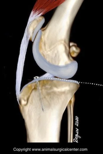
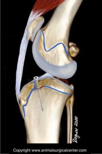
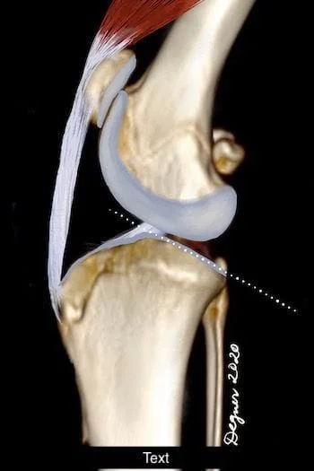
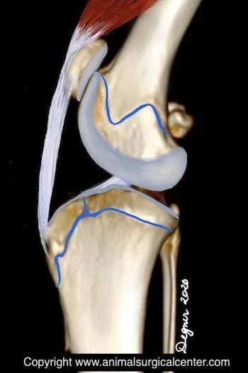
Below is a preoperative and postoperative dynamic TPLP radiographs. Take note of the 26 degree slope at the time of surgery; the cancellous screw is placed at the cranial aspect of the tibial plateau. Four months later the tibial slope has decreased to about 4 degrees.

Aftercare
Typically patients receiving the dynamic TPLP have a fairly low morbidity rate associated with the procedure. Postoperative pain appears to be fairly low, as minimally invasive techniques are used to perform the procedure; therefore, these patients can return home the same day as the procedure is performed. Exercise is limited to leash walks for about 2 months. Typically, this is the period of time during which the lameness resolves. To evaluate the effect of the procedure, radiographs of the tibia are taken initially at 8 weeks after surgery, and then every 1 to 2 months until the dog has matured.
Benefits of the dynamic TPLP
Advantages of this technique include short recovery from surgery, minimal pain, and good clinical results. If indicated, the procedure can also be safely performed bilaterally.
