

Key Points
A perineal hernia is a condition that occurs in both dogs and cats in which there is an abnormal displacement of pelvic and/or abdominal organs (small intestine, rectum, prostate, bladder, or fat) into the region around the anus called the perineum
A perineal hernia is most successfully treated using the internal obturator muscle flap technique
Castration is always performed at the same time as the perineal hernia surgery so that the prostate will shrink, thus minimize straining during bowel movements
Success is highly dependent on the training and experience of the surgeon
Downloadable forms/information for clients for initial visit:
History form - initial consultation for perineal hernia
Client education handout for perineal hernia
Discharge instructions for perineal hernia postop care
Recheck Evaluations
History form - 2 week Telemedicine recheck
History form - 6 week in hospital recheck
Downloadable forms for referring veterinarians:
Overview video describing treatment of perineal hernias in dogs:
Anatomy


The pelvic diaphragm is a set of muscles that are attached to each other and surround a hole called the anus. This diaphragm is a dam that keeps the internal organs such as bowel, prostate and bladder in place.
The pelvic diaphragm consists of the levator ani, coccygeus and external anal sphincter muscles. The above far right illustration above shows the pertinent structures of the pelvic region: C = coccygeus muscle; L = levator ani muscle; AS = anal sphincter; IO = internal obturator muscle; ST = sacrotuberus ligament; P = base of the penis
Take note in the illustration above left showing the sciatic nerve that runs along the side of the pelvic canal and then extends down the hind limb. A branch from the nerve, the caudal rectal nerve, controls the anal sphincter which is important for fecal continence. In the above middle illustration, take note of the caudal gluteal artery/vein which sends a branch, to the muscles in the perineum including the anal sphincter.
Perineal hernia – definition and clinical signs
A perineal hernia is a condition that occurs in both dogs and cats in which there is an abnormal displacement of pelvic and/or abdominal organs (small intestine, rectum, prostate, bladder, or fat) into the region around the anus called the perineum. This condition occurs secondary to a weakening of the muscles, which form the pelvic diaphragm. Signs of this condition include straining to urinate or have bowel movements, constipation, and swelling around the anal region.
Causes of Perineal Hernias
The reasons for development of this disease are not completely understood. The vast majority of cases occur in intact male dogs that are middle-aged or geriatric. The most probable cause is prostate enlargement due to the animal not being neutered. Straining due to an enlarged prostate weakens the pelvic diaphragm. Other theorized causes of perineal hernias include anatomic factors, hormonal imbalances, damage to the nerves of the pelvic diaphragm, and straining due rectal disease.
Diagnosis
The condition is easily diagnosed by digital rectal palpation during a physical examination. X-rays, CT scan or ultrasound may be required to further define the hernia and your pet's overall health.
The Day of Surgery
In preparation for surgery, your pet should be fasted starting at 10 PM the night before surgery; however, water does not need to be with held. To help prevent heartburn after surgery, a single dose of Pepcid AC (10 mg tablet per 20 pounds of body weight) should be administered at 6 AM at home on the day of surgery. Our anesthesia and surgical team will prescribe a pain management program, both during and after surgery, that will keep your companion comfortable. This will include a combination of general anesthesia, injectable analgesics, and oral analgesics.
Surgery


A perineal hernia does not require emergency surgery, however, if the bladder is located in the hernia, emergency treatment may be needed, as the pet may not be able to urinate.
A perineal hernia is most successfully treated using the internal obturator muscle flap technique. This surgical procedure creates a new pelvic diaphragm with the transposed muscle flap. The internal oburator muscle is an external rotator muscle of the hip and can be safely sacrificed with no ill effect to use of the limb.
Other procedures that may be performed include the following:
Colopexy, a procedure in which the colon is tacked to the left body wall and helps to prevent pressure on the repaired hernia.
Cystopexy, a procedure in which the bladder it tacked to the right body wall.
Ductus deferensopexy, a procedure in which the ductus deferens (a cord that goes to the testicle) is attached to the body wall to help prevent the prostate from herniating
These procedures are never used as a primary treatment, rather are always used in conjunction with the obturator flap procedure.
Castration is always performed at the same time as the perineal hernia surgery so that the prostate will shrink, thus minimize straining during bowel movements.
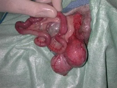
Below is the hind end of a dog that has a severe perineal hernia (photo below left). When the hernia is opened, small intestine, bladder and prostate were found to be in the hernial sac (photo below right). These organs were placed back into the abdomen and the hernia was repaired.

Convalescence period
By 10 to 14 days after the surgery most of the swelling at the level of the surgery will have resolved. Some straining during bowel movements is expected and usually will abate in 7 days. By 6 to 8 weeks after surgery, complete healing has taken place.
Success
Surgery is successful 85 to 90% of the time. Success is highly dependent on the training and experience of the surgeon. In my opinion this type of case usually should be referred to a specialist for surgery. Failure of the surgical procedure results in recurrence of the hernia. A second procedure would be needed to repair the hernia again.
Potential complications
Infection is an uncommon complication of this procedure. Straining to have a bowel movement, can be due to irritation and inflammation of the rectum, which is adjacent to the surgical site. Adding Metamucil to your pet’s food after the surgery will soften the stools so that bowel movements will occur more easily. Fecal incontinence is more common if your pet has hernias on the left and right sides. The cause of the incontinence is due to weakening of the anal sphincter. With this type of problem, stool may accidentally fall out of the anus when the pet is exited, or while barking. In most cases, this is a temporary problem. Urinary incontinence is usually not seen unless the bladder has been chronically located in the hernia. This is due to stretching of the nerves of the bladder and usually resolves with time. Profound lameness of one or both hind limbs can be present and is due to snagging the sciatic nerve (see illustration showing path of the sciatic nerve) during the surgical procedure; if present, emergency surgery should be done to remove the offending suture(s). Anesthetic death is an uncommon complication with routine perineal hernia repair.
Postop care
Stool softeners should be administered to the pet to help minimize the straining and constipation. If your pet has a significant amount of straining a medication called Proctofoam will be prescribed; it has a local anesthetic and a steroid in it. It should only be needed for a couple of days. Pain medication will be prescribed to minimize discomfort. Licking the incision can be prevented by using an Elizabethan collar. Sutures do not need to be removed as these usually are internal and will dissolve with time. During the first 3 weeks after surgery, activity is restricted to short leash walks outside. Running, jumping, or rough play is forbidden. Gradually increasing the activity, following the third week after surgery, allows for a safe recovery.
