

Key Points
OCD of the shoulder is a disease that causes lameness in young large breed dogs
Arthroscopic removal of the OCD flap is less painful than the traditional surgery and results in a quicker recovery
Arthroscopy also allows examination of the entire joint which greatly improves the ability of the surgeon to remove all loose cartilage fragments
Prognosis following surgery is excellent for return to function of the limb
Downloadable forms/information for clients for initial visit:
Client initial consultation history sheet
Shoulder OCD client education handout
Discharge instructions for shoulder surgery postop care
Downloadable forms/information for clients for 2 and 8-week recheck visits:
Shoulder surgery discharge instructions
Downloadable forms for referring veterinarians:
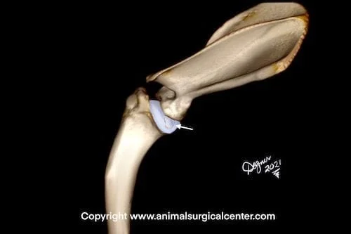
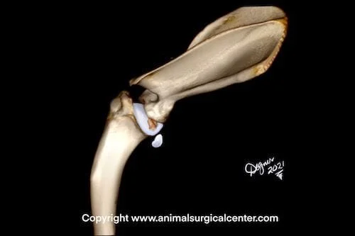
OCD affects young, large-breed dogs and causes lameness of the affected fore limb. OCD is a condition in which a piece of cartilage becomes partially or fully detached from the surface of the joint. Sometimes the cartilage flap becomes detached from its bed and falls into a pocket of the joint (called the cul de sac of the shoulder). These loose fragments can cause inflammation of the lining of the joint and cause pain. Occasionally a fragment of the OCD flap will migrate down the sheath of the biceps tendon (which communicates with the shoulder joint) and causes inflammation of the tendon’s sheath called biceps tenosynovitis. For this reason, surgery should not be delayed.
About 50% of the dogs have osteochondrosis of both shoulders. Dogs usually have a lameness of only one of the limbs, even if both shoulders have a visible osteochondrosis lesion as seen on radiographs. Sometimes the lameness will shift from one leg to the other. If we see OCD on the radiographs on both shoulders, we recommend arthroscopy on both shoulders.
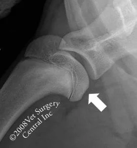
Above right, is a radiograph of a dog’s shoulder that has an OCD lesion of the head of the humerus. Take note of the dent in the surface of the bone (labeled with the white arrow)
Arthroscopic Surgery
During arthroscopic surgery a small incision is made in the skin. To confirm that the needle is in the joint, a syringe is attached to the needle and aspirated. If joint fluid is retrieved, the surgeon is certain that the needle is within the confines of the joint. Next, the surgeon will inject sterile fluid (lactated Ringer’s solution) into the joint which will distend the joint. This makes it easier for the surgeon get the scope into the joint. The surgeon then passes a hollow tube called a cannula into the joint
The telescope of the camera is inserted into the cannula and a rubber hose is attached to the cannula so that fluid can be infused into the joint during the procedure. The infusion of fluid will keep the field of view clear for the surgeon during the arthroscopic surgery and will flush out small fragments of loose cartilage.
An instrument is inserted into the shoulder joint and the OCD flap is grasped and is pulled out of the joint. In some cases the flap is taken out in pieces and other cases the flap can be removed in one piece. In this case, the OCD flap is torn into smaller fragments and removed in multiple steps. Play video. The surgeon will examine the front of the shoulder and confirm a diagnosis of OCD. Take note of the large cartilage flap that is seen in this joint.
Traditional surgery (open surgical approach)
During an open surgical approach to the shoulder joint, a 2 inch incision is made and the muscles are just separated along their natural anatomical planes. Because we do not actually cut any muscles, tendons, or ligaments during the surgery, the recovery period after surgery is relatively short. The disadvantage of the open approach is that a less thorough examination of the joint and its associated structures can be made and loose pieces may be left behind in the front part of the shoulder joint.
Convalescent period
Arthroscopic surgery. The recovery from surgery is faster if the procedure is done arthroscopically versus with an open surgical approach. Most dogs are bearing weight on the limb at the time of release from our hospital. Within two weeks the lameness generally is very mild.
Traditional surgery. Within a couple of days after the surgery most pets will bear some weight on the operated limb. By two weeks after surgery many of the operated dogs have mild to moderate lameness. By 8 weeks after surgery, your pet should be using the limb normally. Recovery can be somewhat variable from one pet to another.
Success
Surgery is about 90% successful. Success is defined as the return of good function of the limb (i.e. lameness resolves). Surgery will not remove the arthritis that is already present in the shoulder. As a result, some pets may have some stiffness of the limb or lameness after very heavy exercise or during weather changes (cool damp conditions). Most dogs that have surgery do not develop significant arthritis after surgery and have no lameness or stiffness. Continued lameness can be also due to biceps tendonitis.
Postop care
During the first 6 to 8 weeks after surgery, activity is restricted to short leash walks outside. Running, jumping, or rough play is forbidden. Gradually increasing the activity, following the eighth week after surgery, allows for a safe return of function of the operated limb.
Potential complications
Following arthroscopic surgery, the most common complication is swelling of the shoulder due irritation of the joint with fluid during the surgery. The swelling typically resolves within 24 to 48 hours after surgery. Uncommon complications include infection, inability to retrieve the OCD fragment thus necessitating conversion to an open surgical procedure (uncommon…I have not needed to convert to open surgery in years), and anesthetic death (very rare). Unresolved or recurrent lameness may be due to biceps tenosynovitis; this can be due to small pieces of the cartilage flap that have become lodged deep within the bursa of the biceps tendon.
Following open surgery, the most common complication following open surgery is the development of a seroma at the level of the surgical incision. A seroma is an accumulation of fluid under the skin. This usually feels like a soft, fluid-filled sac. The cause of this complication is excessive movement of the tissues at the level of the shoulder (this is just the nature of the area). Restricting activity after surgery will minimize seroma formation. Seromas usually resolve without any treatment. Other uncommon complications include infection, unresolved lameness, and anesthetic death.
