Key Points
- The radius is the main weight-supporting bone; the ulna bone supports very little weight.
- Small breed dogs have a poor blood supply to the lower fourth of the radius bone, therefore it is more susceptible to being fractured; also healing of the fracture can take longer than other bones in the body.
- Surgical fixation of the fractures with plates and screws or with an external skeletal fixator is usually needed for a successful outcome.
Initial visit- downloadable forms/information for clients:
History sheet - Client initial consultation history
Client education handout - radius/ulnar fractures
Discharge instructions for postop care with bandage - dog
Discharge instructions for postop care with cast/splint - dog
Discharge instructions for postop care with bandage - cat
Discharge instructions for postop care with cast/splint - cat
2-week recheck visit - downloadable forms/information for clients:
History form - 2 week postop (Telemedicine evaluation) - dog and cat
Postop care instructions 2-week fracture repair recheck - cat
Postop care instructions 2-week fracture repair recheck - dog
8-week recheck visit - downloadable forms/information for clients:
History form 8-week fracture recheck (in hospital) - dog and cat
Postop care instructions 8 week fracture recheck - dog
Postop care instructions 8 week fracture recheck - cat
Downloadable forms for referring veterinarians:
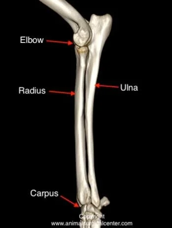
Anatomy
The fore limb has two bones between the wrist (carpus) and the elbow joint called the radius and ulna bones. The radius is the main weight-bearing bone; the ulna bone is a minor weight-bearing bone. Small breed dogs have a poor blood supply to the lower fourth of the radius bone; therefore, the radius is susceptible to being fractured. Small breed dogs also may have slow bone healing of a radius bone fracture. Large breed dogs have a much better blood supply to this region, therefore a very substantial force needs to be applied to the bone for a fracture to occur. When the radius bone fractures, the ulna usually fractures too.
Cause of fracture
In small breed dogs, landing on the front limbs from a fall is the most common cause of fracture of the radius (example: dog falls out the owner's arms or jumping off a sofa). In large breed dogs, substantial trauma is needed to break the bone, such as being hit by a car. Gun shot injuries will result in an open fracture. This results in dirt and hair being driven into the tissues and can result in infection of the soft tissues and bone.
Preparation for surgery
Your pet should be fasted prior to surgery, as instructed by the surgical team. Water is usually permitted up to the time of admission to our hospital. You should inform the surgical team of any medications that your pet is currently receiving and of any pertinent medical alerts (allergies to food, seizures, drug insensitivities, etc). Just prior to surgery, your pet will receive a sedative, have an intravenous catheter placed for the administration of intravenous fluids and intravenous medications, be induced under general anesthesia with medication(s), and have a breathing tube (endotracheal tube) placed to allow delivery of oxygen and gas anesthetic agent. The surgical site will be clipped and cleansed with an anti-septic solution in preparation for surgery. While under general anesthesia, your pet’s breathing will be assisted with a ventilator and vital parameters such as heart rate, respiratory rate, core body temperature, blood pressure, oxygenation of the blood (pulse oximetry), exhaled/inhaled carbon dioxide (capnography), and heart rhythm (EKG) will be monitored to ensure the pet’s well being. Pain will be controlled both during and after surgery with analgesics (pain-controlling medication). Please note that each surgical and anesthesia team may elect to choose a different, but effective analgesia protocol, pending the needs of the patient.
Surgery
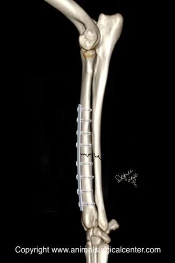
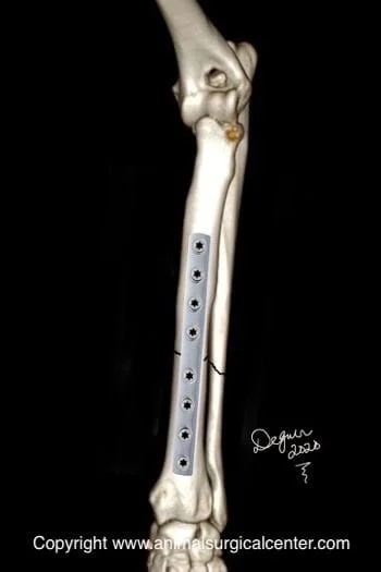
The best option to stabilize radial/ulnar fractures is with a bone plate and series of screws. This treatment has the least aftercare for the client and a good prognosis for uneventful bone healing. The plate is commonly placed on the top side (dorsal) of the radius bone. Because the ulna bone does not have significant load-bearing, it usually is not repaired with a metal plate. Below are illustrations showing the radius/ulnar fractures (front view) with a bone plate applied to the bone and a side view showing the bone plate and screws.

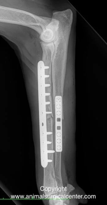
In larger dogs, in which there is comminution of the bone (multiple fractures), the ulna bone may also require stabilization with a plate and screws.
In some cases, the fracture can be done as a minimally invasive procedure with a relatively small incision made at the top and at the bottom of the radius bone. A bone plate is slid over the bone and then secured with 2 or 3 screws at each end of the bone. Minimally invasive bone fracture repair usually results in quicker bone healing.
An external fixator consisting of multiple pins that penetrate the skin and bone and an external connecting bar that runs parallel to the bone may be used in cases in which a contaminated open wound is also present at the fracture site. This apparatus can be also used for highly comminuted fractures of the radius bone. As the bone heals, the external skeletal fixator is removed, usually in multiple stages.
A single pin placed in the marrow cavity of the radius bone to hold the fracture together is an old technique and is considered undesirable, as the risk of poor bone healing is significant.
Use of a cast to stabilize the bones frequently results in a nonhealing fracture, therefore this form of treatment is considered an unacceptable option in most cases. Green-stick or incomplete fractures may be considered for casting.
Aftercare
A bandage is kept on for 5 to 10 days after surgery in most cases. In some cases, a splint or cast will also be applied to protect the internal surgical repair during the bone healing phase. If an external fixator has been placed, dressing changes will need to be completed daily until the skin has healed around the pins. The incision should be checked for signs of infection until healing has taken place. Exercise must be restricted until the fracture has healed (based on radiographs). Radiographs (x-rays) are taken in 8 weeks after the surgery. However, if the patient is only a few months of age, radiographs may be made earlier, as bone healing occurs quickly.
Potential Complications
Complications may include, infection, nonhealing of the fracture and breakage of the surgical implants before healing of the fracture has occurred. Catastrophic implant failure/breakage is usually is associated with over activity during the healing process. Stress protection or stress shielding is an uncommon complication of plating a bone fracture in which the bone becomes so fragile (osteoporosis) that a fracture of the bone at the end of the plate may occur. In this condition, the bone relies on the metal for its strength and the body resorbs calcium from the bone. Removal of the bone plate/screws after healing occurs can prevent this complication. Your pet's surgeon can make a recommendation when it is safe to remove surgical implants.
If a growing pet has damage or fracture of a growth plate of the radius or ulna, the limb may become twisted or bent as the pet grows. This would require a second surgery to straighten the limb out.
Success
Most patients will have uneventful healing of the fractures within 8 weeks after surgery. In some cases, the ulna bone does not heal together or may partially resorb in the area of the fracture; however, this does not seem to affect the clinical outcome.
Case examples of bad ways to fix radial/ulnar fractures:
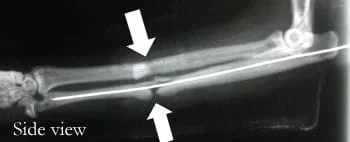
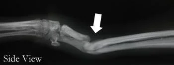
Case 1: Poodle, 1.5 yrs old, spayed female
History: dog sustained fracture after jumping out of owner’s arms, a veterinarian treated the fracture initially using splint. Below is a side view and and front view radiographs taken 3 months of splinting (arrow points to fracture in bone). The result was a bent, nonfunctional limb and fracture which did not heal. To prevent such a complication, a plate and screws should have been used to stabilize the fracture.

Case 2: Domestic short haired cat, 4 years old, spayed femal
History: cat was hit by a car and susteined fractures of the radius and ulna
A veterinarian repaired this fracture using a single pin in the ulna and did not stabilize the radius. Because the radius is the weight-bearing bone, it is imperative that this bone stabilized with a plate, not just another pin. Pinning this bone does not overcome rotation at the fracture site, therefore the chance of healing is poor. This cat developed a nonunion of the fracture. Best treatment again would be stabilization with a plate and screws.