

Key points
- Femoral fractures is almost all cases should be stabilized with surgery
- Femoral fractures left to heal on their own may either not heal at all or they may heal in a maligned fashion rendering the limb nonfunctional or result in hip arthritis
- The most common form a stabilization used at Animal Surgical Center of Michigan include plate/screws or interlocking nail
Initial visit- downloadable forms/information for clients:
History sheet - Client initial consultation history
Client education handout - femoral fractures
Discharge instructions for postop care - dog
Discharge instructions for postop care - cat
2-week recheck visit - downloadable forms/information for clients:
History form - 2 week postop (Telemedicine evaluation) - dog and cat
Postop care instructions 2-week fracture repair recheck -cat
Postop care instructions 2-week fracture repair recheck -dog
8-week recheck visit - downloadable forms/information for clients:
History form 8-week fracture recheck (in hospital) - dog and cat
Postop care instructions 8 week fracture recheck - dog
Postop care instructions 8 week fracture recheck - cat
Downloadable forms for referring veterinarians:
Basic facts and anatomy
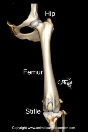
A fracture is synonymous with a broken bone. The bone between the hip and the stifle (knee) joints is called the femur bone (see illustration right). The top of the femur bone has a head and neck and a greater trochanter. The bottom of the femur bone consists of the medial and lateral condyles. The femur bone is very well muscled which provides a very good blood supply to the bone. The hind limb is innervated by the sciatic and femoral nerves. The sciatic nerve runs along the backside of the femur bone and may be injured by a sharp broken femur bone, whereas the femoral nerve is less likely to sustain significant injury. Puppies have growth plates at the ends of the femur bones from which the bone grows. Growth plates are susceptible to developing fractures in immature animals.
Causes of fractures
The most common cause of a femoral fracture is trauma such as being struck by a motorized vehicle. Gunshot injuries not only will fracture the bone, but also result in a dirty open wound. This could potentially result in infection and delayed healing of the bone. If the fractured bone is sharp it may penetrate through the skin and result in infection of the bone. If the pet sustains a fracture without any known trauma, there may be an underlying disease that has weakened the bone such as a nutritional deficiency. Foods that have too much phosphorus and too little calcium or too much vitamin A will make the bones weak. Some animals have an inherited collagen defect that weakens the bones, resulting in bone fractures with minimal trauma. Bone cancer also can weaken the bone and predispose the pet to a spontaneous fracture.
Preparation for surgery
Your pet should be fasted prior to surgery, as instructed by the surgical team. Water is usually permitted up to the time of admission to our hospital. You should inform the surgical team of any medications that your pet is currently receiving and of any pertinent medical alerts (allergies, seizures, drug insensitivities, etc). Just prior to surgery, your pet will receive a sedative, have an intravenous catheter placed for the administration of intravenous fluids and intravenous medications, be induced under general anesthesia with medication(s), and have a breathing tube (endotracheal tube) placed to allow delivery of oxygen and gaseous anesthesia. The surgical site will be clipped and cleansed with an anti-septic solution in preparation for surgery. While under general anesthesia, your pet’s breathing will be assisted with a ventilator and vital parameters such as heart rate, respiratory rate, core body temperature, blood pressure, oxygenation of the blood (pulse oximetry), exhaled/inhaled carbon dioxide (capnography), and heart rhythm (EKG) will be monitored to ensure the pet’s well being. Pain will be controlled both during and after surgery with analgesics (pain-controlling medication). Please note that each surgical and anesthesia team may elect to choose a different, but effective analgesia protocol, pending the needs of the patient.
Surgery
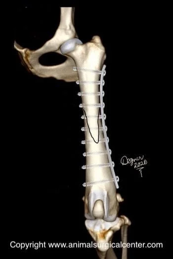
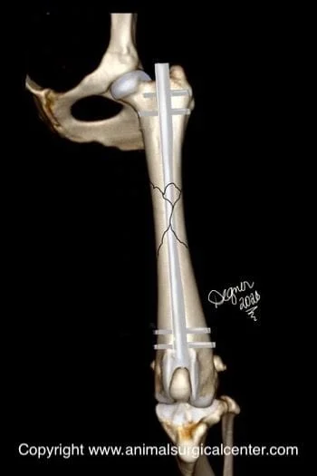
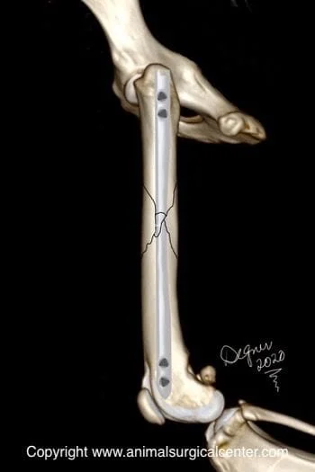
Two-piece fractures of the femur are commonly stabilized with a bone plate and multiple screws (see below). Femoral fractures that are best amenable to plating are those that can be reconstructed, have bone fragments that can share the weight load (i. e. plate is not the only supporting structure). If these features cannot be achieved, there is increased risk for the plate to break in the postop period, especially in a very active, rambunctious pet. The addition of a pin down the marrow cavity, in the form of a pin-plate repair, will help take stress off the metal plate.

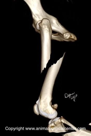
If the bone is broken in multiple pieces, called a comminuted fracture, an interlocking nail is typically the fixation apparatus of choice (see below). Both of these treatment options provide the least aftercare for the client and a successful outcome in most cases. Femur bone fractures cannot be casted or successfully splinted. In addition, this form of treatment usually will result in a failure of the bone to heal or inappropriate healing of the bone (misaligned bone).

Home care
After surgery, you can continue to give your pet a prescribed pain reliever to minimize discomfort. It’s also extremely important to limit your dog’s activity and exercise level during this post-operative period. Rehabilitation exercises can be done at your home or if you choose, by professionally trained therapists at an animal rehabilitation center. Rehabilitation therapy should be continued until your dog is bearing weight well on the operated limb (typically 2 weeks after surgery). Detailed instructions will be given to you after the surgery. The surgeon will monitor the healing process with two follow-up exams. The first is scheduled at two weeks after the surgery and the second is at eight weeks after the surgery. By 8 to 10 weeks after surgery, most dogs are fully weight bearing on the operated limb, although exercise should be limited during the first three months after the procedure.
Results
Surgical repair of a fractured femur with a plate and screws or I-loc offers multiple benefits including a faster recovery, earlier use of the limb after surgery, better chance to return to athletic activity, less risk of a second surgery being required, and better range of motion of the joints above and below the fracture. Uncommon complications include infection, nonhealing of the fractures, breakage of the metal plate, osteoporosis of the bone, bone cancer induced by metal implants, and sciatic nerve damage.
