

Key Points
Dislocation of the kneecap is painful and results in lameness
Evaluation of limb alignment may be recommended, especially in large breed dogs to assess the need to correct this; failure to address this can result in increased failure rates
Concurrent cruciate ligament injury may be present in dogs that have a dislocating kneecap, therefore this may also need to be addressed
Prognosis generally is very good – the higher the grade of the patellar luxation, the greater the failure rate unless more corrective surgeries are done
Initial visit- downloadable forms/information for clients:
History sheet - Client initial consultation history
Client education handout - Patella Luxation
Discharge instructions for postop care - dog
Discharge instructions for postop care - cat
2-week recheck visit - downloadable forms/information for clients:
History form - 2 week postop (Telemedicine evaluation)
Postop care instructions 2-week recheck - dog
8-week recheck visit - downloadable forms/information for clients:
History form 8-week recheck (in hospital)
Postop care instructions 8 week fracture recheck
Downloadable forms for referring veterinarians:
Anatomy
The patella also known as the kneecap normally rides in the trochlear groove which is located at the bottom of the femur bone. The quadriceps muscles are attached to the top of the patella. The patellar ligament connects that patella to the tibia on a bony protrusion called the tibial crest. Ligaments on the sides of the patella prevent it from dislocating out of the groove. The illustration right shows the patella dislocated out of the groove.
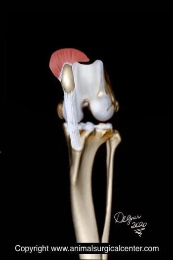
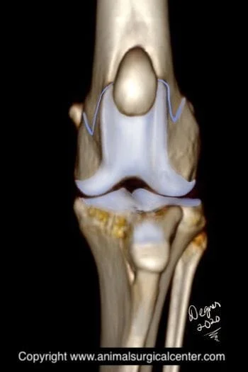
The patella (commonly known as the kneecap) normally rides in a groove at the bottom of the femur at the level of the knee joint in a groove called the trochlear groove; Fig 1 and 2 show a front view of the knee joint; Fig 1 demonstrates the patella in the groove, where as Fig 2 demonstrates the knee cap dislocated out out of the groove (knee is flexed in this illustration).
Patellar luxation is caused by congenital abnormality usually at the level of the hip joint and results in abnormal forces on the kneecap, which cause it to eventually ride outside of the groove. The groove becomes very shallow and the attachment of the ligament of the patella may be malpositioned on the tibia bone. If the patellar luxation occurs in immature animals, the tibia and femur bones become twisted.
Clinical signs
Lameness
Intermittent skipping gait
Pain
Stiffness of the hind limb
Some pets show only a single sign, whereas others show many signs of the condition
Failure to treat the condition could lead progressive debilitating arthritis of the joint
Preparation for surgery
The pet should be fasted prior to surgery, as instructed by the surgical team. Water is usually permitted up to the time of admission to the hospital. The surgical team should be informed of any medications that your pet is currently receiving. Just prior to surgery, your pet will receive a sedative, have an intravenous catheter placed for the administration of intravenous fluids and intravenous medications, be induced under general anesthesia with medication(s), and have a breathing tube (endotracheal tube) placed to allow delivery of oxygen and gaseous anesthesia. The surgical site will be clipped and cleansed with an anti-septic solution in preparation for surgery. While under general anesthesia, the pet’s breathing will be assisted with a ventilator and vital parameters such as heart rate, respiratory rate, core body temperature, blood pressure, oxygenation of the blood (pulse oximetry), exhaled carbon dioxide (capnography), and heart rhythm (EKG) will be monitored to ensure the pet’s well being. Pain will be controlled both during and after surgery with analgesics (pain-controlling medication). Please note that each surgical and anesthesia team may elect to chose a different, but effective analgesia protocol.
Surgery
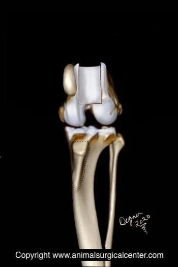
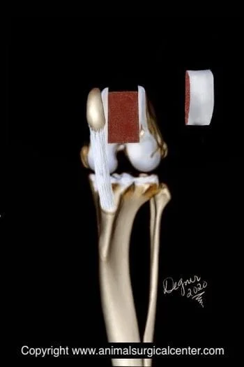
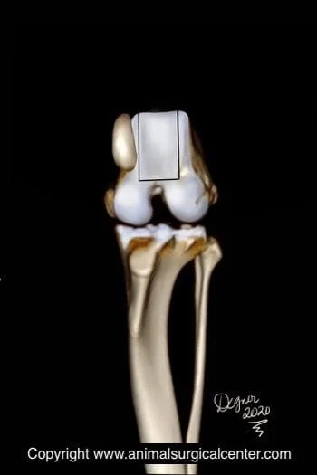
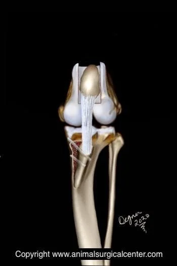
During the surgery, the surgeon will make an assessment as to which procedures are best suited to correct patellar lunation in your pet. If the trochlear groove is shallow, it will be deepened. If the cartilage has been damaged, the surgeon likely will perform an excisions trochleoplasty, however, if the cartilage is in good condition, a block trochleoplasty will be performed. In this procedure two vertical and on horizontal bone cut will be made (see below left). A block of cartilage and bone is lifted off the bed (see below middle). Additional bone is removed from the raw bone bed and the cartilage/bone plate is replaced creating a deep groove for the patella to ride in (see below right). The benefit of the block osteotomy is that it uniformly deepens the groove from top to bottom and preserves the cartilage that the patella rides on.
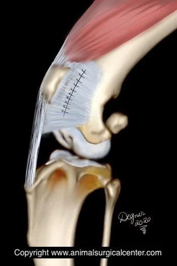
If the attachment of the patellar ligament to the tibia, called the tibial crest, is in the wrong position, it is repositioned. This is done by creating a cut in the tibial crest and reattaching the bone in a position so that the patella is realigned within the trochlear groove. Pins are used to fasten the bone in place (see illustration right). The pins usually do not need to be removed unless they migrate out of position or a bubble of fluid (seroma) develops over the end of the pin and does not resolve after a few months.
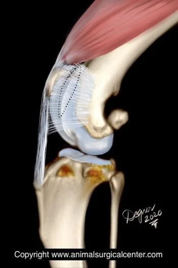
The soft tissues along the side of the patella usually are stretched and are tightened to provide additional support to keep the patella in the trochlear groove.
The femur bone may be twisted in some dogs which worsens the condition of the luxating patella. Dogs that have a greater than a 14 degree bowing (varus) of the thigh bone should have this surgically corrected. CT scan is usually made in large breed dogs to check for this problem and a 3D printed model can be made for surgical planning purposes.
How to care for your pet after surgery
Surgery is only one part that will lead to a successful outcome. The second part is on the pet owner. Prescribed medications should be given to make sure that the pet's pain is well controlled, antibiotics to prevent infection and sedatives if the pet is hyperactive. Exercise must be restricted. Walking on the limb is not going to hurt the operated limb, but explosive activity is dangerous. Rehabilitation therapy is done at home and the recovery process may also be accelerated with the help of a rehabilitation therapist. Below is a video that demonstrates range of motion exercises that should be done after surgery. Your pet must not lick the incision, therefore an Elizabethan collar should be worn for the first 2 weeks after surgery.
Convalescence
By 10 to 14 days after the surgery, your pet should be touching the toes to the ground at a walk
By 2 to 3 months after surgery your pet should be using the limb well
If your pet does not follow a normal progression of recovery, the surgeon should be notified
Prognosis
Surgery has approximately a 90% success rate. Success is defined as the return of good function of the limb
Unfortunately surgery will not remove the arthritis that may already be present in the knee. As a result, your pet may have some stiffness of the limb in the mornings or after laying down for a nap. In addition, your pet may have some lameness after heavy exercise
By having the surgery done earlier, the chance of developing significant arthritis is decreased
Dogs that have a higher grade of patellar luxation may have increased risk for reluxation of the patella
Large breed dogs that have patellar luxation may have increased risk for reluxation of the patella if a corrective femoral osteotomy is not performed
Potential complications
In the event of an uncommon reaction to anesthesia, death may occur
Infection of the surgical site, although not common, can occur
Pin migration or pin breakage
Seroma formation over the pins
If exercise is not minimized for 8 weeks after the surgery, breakdown of the repair may occur, thus requiring a second surgery
