

Key Points
- Cranial cruciate ligament rupture is the most common orthopedic condition of dogs and affects all breeds. Occasionally cats and ferrets develop the same problem
- The cranial cruciate ligament is one of the main stabilizing structures of the stifle joint
- The lateral imbrication technique has good outcomes in small breed dogs, but is not the best option in large breeds
Initial visit- downloadable forms/information for clients:
History sheet - Client initial consultation history
Client education handout - Lateral Imbrication
Discharge instructions for postop care - dog
Discharge instructions for postop care - cat
2-week recheck visit - downloadable forms/information for clients:
History form - 2 week postop (Telemedicine evaluation)
Postop care instructions 2-week recheck - dog
Postop care instructions 2-week recheck - cat
8-week recheck visit - downloadable forms/information for clients:
History form 8-week recheck (in hospital)
Postop care instructions 8 week fracture recheck - dog
Postop care instructions 8 week fracture recheck - cat
Downloadable forms for referring veterinarians:
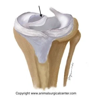
Anatomy

The illustration right is a front view of the stifle joint; note the medial meniscus (M), lateral meniscus (L), cranial cruciate ligament (Cr), and caudal cruciate ligament (Ca).
Cranial cruciate ligament rupture
Cranial cruciate ligament rupture is the most common orthopedic condition that we treat and affects all breeds of dogs, and occasionally cats and ferrets. The ligament may undergo partial tearing over a period of months, or may suddenly rupture during normal physical activity. Most dogs are middle-aged or older when the rupture occurs, however some breeds such as Labrador Retrievers, Rottweilers, and Mastiffs may develop partial or complete tears when they are puppies. The cause is unknown, but conformation of the limbs and genetics may play a role. Partial ligament tears may be difficult to diagnose and frequently occur in both legs at the same time. When the ligament tears, the stifle becomes unstable. The femur and tibia bones that form the joint then rub back and forth on each other (termed “drawer movement”). This results in pain due to stretching of the joint capsule, potential damage to the meniscal cartilage, and inflammation of the joint (called arthritis). In about half of the patients that we operate, the meniscal cartilage on the inner side of the joint (medial meniscus) has been torn and the damaged portion must be removed.
Candidates for the lateral fabellar surgery
This technique is used most commonly for small dogs and cats. Large breeds of dogs have a better outcome when the tibial plateau leveling osteotomy (TPLO) is performed. We have noted that dogs that have a steeply-sloped tibial plateau should receive the TPLO instead of the lateral imbrication technique regardless of their size. The lateral imbrication technique cannot overcome the forces on the stifle joint caused by a steep slope and frequently the nylon band will tear or loosen in the postop period. All dogs that are going to have cruciate surgery should have a correctly positioned x-ray taken to measure the slope of the tibia so that an informed decision can be made on the appropriate type of surgery that should be performed. In my experience dogs that have a steep tibial slope (especially large breeds) do much better with the TPLO surgery. This may not be an important factor in small breeds even with a steep tibial slope, but the clients should be aware of the fact that a steep tibial slope will put much greater force on the nylon bands, therefore they may break.
Surgery
The stifle is surgically opened to examine the inside of the joint. The torn ends of the cruciate ligament are removed and the medial and lateral meniscus cartilages are examined for tears. If the medial meniscus is not torn, a prophylactic release of the caudal (back) pole of the meniscus may be performed (surgeon preference) in order to help prevent a tear in the future.
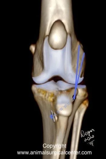
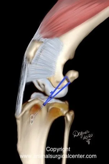
Heavy suture material (monofilament nylon) is passed from the lateral fabella to the tibial crest and tied in order to eliminate joint instability (drawer movement). With time, scar tissue develops around the stifle joint which helps to stabilize the joint. The build up of scar tissue may also decrease the range of motion of the joint. An alternative to the using lateral fabellar technique is the tibial plateau leveling osteotomy. This is an excellent technique for all size dogs, highly athletic dogs (hunting, agility), or show dogs. Some surgeons prefer this technique over the lateral fabellar technique, as these patients recover quicker and develop less degenerative arthritis of the stifle. The illustrations right shows the implanted nylon bands which stabilize the joint. The blue lines represent the nylon bands that are passed around the fabella bone (which sits on the back of the femur bone), and through a hole that has been drilled in the front part of the tibia bone. Some surgeons use a metal button on the medial tibia to anchor the the bands to the tibia and other surgeons pass the band through the fat pad (surgeon's preference). The nylon bands pull the femur back into place on the tibia.
Contralateral cruciate tears
About half of the dogs will also tear the cruciate ligament in the opposite limb. These dogs frequently have arthritis in the knee joint even before the tear in the cruciate is obvious on physical examination, but early changes such as mild joint swelling may be detected with x-rays. An x-ray may be recommended to predict if the opposite stifle joint is going to develop a tear.
Expected convalescent period
By 2 weeks after the surgery your pet should be touching the toes to the ground at a walk. By 8 weeks the lameness should be mild to moderate. By 6 months after the surgery your pet should be using the limb well.
How to care for your pet after surgery
Surgery is only one part that will lead to a successful outcome. The second part is on the pet owner. Prescribed medications should be given to make sure that the pet's pain is well controlled, antibiotics to prevent infection and sedatives if the pet is hyperactive. Exercise must be restricted. Walking on the limb is not going to hurt the operated limb, but explosive activity is dangerous. Rehabilitation therapy is done at home and the recovery process may also be accelerated with the help of a rehabilitation therapist. Below is a video that demonstrates range of motion exercises that should be done after surgery. Your pet must not lick the incision, therefore an Elizabethan collar should be worn for the first 2 weeks after surgery.
Success rates
With the extracapsular technique, about 85% of the cases are significantly improved from their preoperative state. With the extracapsular technique, we can expect that 50% of these dogs will have some degree of lameness, whether it is mild or intermittent following heavy activity. On the other hand about 50% regain normal function of the limb. Even though this surgery may not return the limb to perfectly normal function, these dogs usually are greatly improved over their condition prior to surgery. The lateral fabellar suture technique will not stop the progression of arthritis that is already present in the joint. As a result, your pet may have some stiffness of the limb in the mornings. In addition, your pet may have some lameness after heavy exercise or during weather changes. To help with stiffness chondroitin sulfate and glucosamine may be given.
Potential complications
Anesthetic death can occur, but is rare. Infection at the surgical site is also uncommon. Sterile reaction to the nylon bands necessitates removal the bands. Premature loosening or breakage of the nylon bands may require a second surgery. If the meniscal cartilages were not found to be damaged at the initial surgery, it is possible that damage may occur at a later date, thus requiring a second surgery. The sign of a meniscal tear is a sudden onset of lameness. Entrapment of the peroneal nerve by the nylon sutures is very painful and can result in permanent functional impairment of limb function.
Postop care
Ice pack the stifle three times daily, 10 minutes per session for the first 2 days to help reduce the swelling and pain. Hotpack the stifle starting on the fourth day after surgery, 10 minutes per session prior to passive range of motion of the joint. The hotpacking will soften the soft tissues prior to the range of motion of the joint and will make it less painful to do the exercise. Passive range of motion of the joint involves flexing and extending the stifle joint, and should be done 10 minutes following the hotpacking. After the range of motion exercises have been completed, a cold compress is applied to the stifle for about 5 to 10 minutes. The rehabilitation therapy should be done until your pet is bearing a significant amount of weight on the limb. During the first two months activity is restricted to short leash walks outside. Running, jumping, and rough play are forbidden. Gradual increasing the activity during the third month after surgery allows for a safe return to function. Unleashed activity can take place after three months.
