

Key Points
Breathing difficulty in a brachycephalic dog or cat is commonly due to
+ Stenotic nares
+ Elongated soft palate
+ Everted lateral ventricles of the larynx
+ The best prognosis following surgery is in pets that are immature
Initial visit- downloadable forms/information for clients:
History sheet - Client initial consultation history
Upper airway examination form
2 wk postop visit- downloadable forms/information for clients:
History sheet
Discharge instructions
8 wk postop visit- downloadable forms/information for clients:
History sheet
Discharge instructions

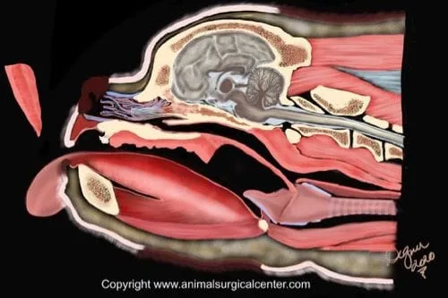
Air is passed from the nares (nostrils), through the nasal cavity, back of the throa, and into the trachea via the larynx.
Definitions:
Nares = nostrils
Nasal cavity = the nose cavity
Turbinates = scroll like bones in the nasal cavity that are lined with mucosa which serve to warm and moisten inhaled air
Mucosa = the skin like tissue that lines the inside of the nose and mouth
Trachea = windpipe
Larynx = voice box
Nasopharynx = airway passage that connects the nasal cavity to the back of the throat called the pharynx
Lateral ventricles = cavities that are located adjacent to the opening of the voice box
Hard palate = roof of the mouth (hard portion) that separates the mouth cavity from the nasal cavity
Soft palate = a soft tissue structure extending from back of the hard palate which separates the back of the throat (pharynx) from the nasopharynx

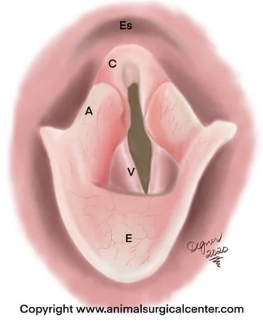
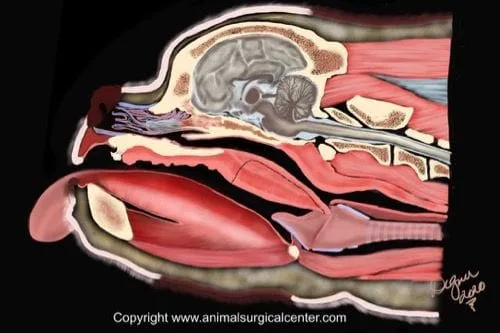
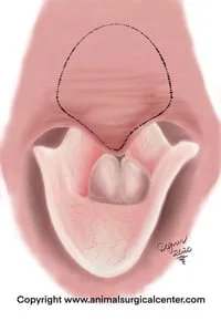
The sagittal cross-sectional view of a brachycephalic dog (see right) shows a compacted nasal cavity, thick tongue and very thick elongated, thick soft palate that extends into the opening of the larynx. The illustration below shows anatomy of the larynx which includes the epliglottis (E), vocal cords (V), Cuneiform process (C) of the arytenoid and the Corniculate process (A) of the arytenoid. The opening of the esophagus (Es) is just above the larynx. The soft palate is not illustrated here.
What is brachycephalic airway syndrome?
This condition is due to a compacted the upper airway which distorts the normal anatomy. The result is that structures that normally fit well within the head and neck are disproportionate, thus the airway is not as open as it ought to be. The soft palate is frequently elongated in brachycephalic breeds (bulldog, Boston terrier, Shar pei, Pug, Himalayan cats, etc) and frequently will occlude the opening of the larynx. In addition, the nares commonly are narrower than normal in such breeds. Secondarily, the lateral ventricles which are normally concave pockets along side of the larynx become everted to form balloon-like structures which further occlude the opening of the larynx. The larynx may also become weakened with time and collapses together, which will further reduces the flow of air from the nose to the trachea. Another anatomical abnormality is an abnormally narrow windpipe called a hypoplastic trachea.
Other anomalies in brachycephalic include aberrant turbinates that partially occlude the upper airway. A thicker than normal tongue which not only can compress the the nasopharynx and restrict nasal air flow, but also obstructs air flow through the mouth during mouth breathing. These dogs also have increased goblet cells within the mucous membranes which secrete excessive mucus.
Some dogs have a poorly functioning muscle called the hypoepiglotticus muscle. This muscle retracts the epiglottis away from the opening of the larynx. In such dogs, even after surgical correction of the brachycephalic upper airway issues, continued breathing issues persist.
Many brachycephalics have chronic esophagitis (heartburn) due to reflux of fluid from the stomach. This could be due to a combination of increased negative pressure within the chest cavity, lower esophageal incompetence, sliding hiatal hernia and potentially esophageal motility disorder. Regurgitation, subsequent aspiration of stomach fluid and development of pneumonia.
Weight gain is common in affected animals due to sleep apnea in brachycephalic breeds. This condition puts the pet in a state of low metabolism due to oxygen depravation. Fat accumulates in the back of the throat which further impairs the breathing.
Signs
Warning signs of brachycephalic airway syndrome include snoring when sleeping, snorting noises when breathing, collapsing when playing, and a gray or purple color of the tongue. Signs are worsen with heat, exercise and excitement. Other secondary signs may include vomiting or regurgitation. Aspiration pneumonia may also be present with this condition, and signs may include coughing, increased respiratory rate, labored breathing, fever, lethargy, and inappetance.
Diagnostic testing
Blood testing including a complete blood count, chemistry panel, and urinalysis should be done to rule out other conditions that may complicate surgery. An examination of the back of the throat is performed under general anesthesia. This is generally performed at the time of the proposed surgery. CT scan of the head through abdomen with the patient lying belly down and the head suspended; ideal imaging is completed with no endotracheal tube in place.
Preparation for surgery
The pet should be fasted prior to surgery, as instructed by the surgical team. Water is usually permitted up to the time of admission to the hospital. One week prior to surgery, omprazole and cisapride are administered to the pet to help reduce the risk of regurgitation and subsequent aspiration pneumonia. The surgical team should be informed of any other medications that your pet is currently receiving. Just prior to surgery, your pet will receive a sedative, have an intravenous catheter placed for the administration of intravenous fluids and intravenous medications, be induced under general anesthesia with medication(s), and have a breathing tube (endotracheal tube) placed to allow delivery of oxygen and gaseous anesthesia. While under general anesthesia, the pet’s breathing will be assisted with a ventilator and vital parameters such as heart rate, respiratory rate, core body temperature, blood pressure, oxygenation of the blood (pulse oximetry), exhaled carbon dioxide (capnography), and heart rhythm (EKG) will be monitored to ensure the pet’s well being. Pain will be controlled both during and after surgery with analgesics (pain-controlling medication). Please note that each surgical and anesthesia team may elect to chose a different, but effective analgesia protocol.
Surgery
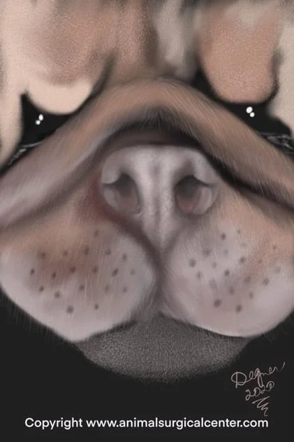
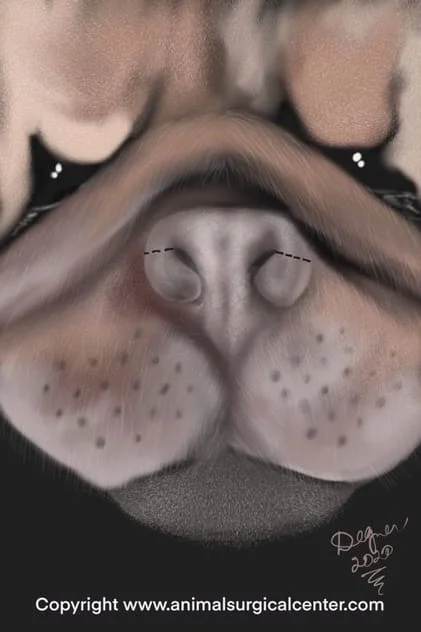
A flow dynamic study in the dog has shown that the stenotic nares contribute the most to air flow obstruction in brachycephalic. Nares are surgically enlarged if they are stenotic. This involves removal of a wedge of tissue from the sides of each nare and cartilage within the nasal entrance. No sutures are needed during this procedure. Raw tissue will heal nicely with one month, leaving both nostrils wide open.

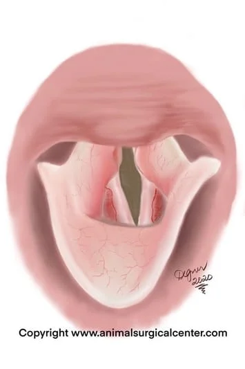
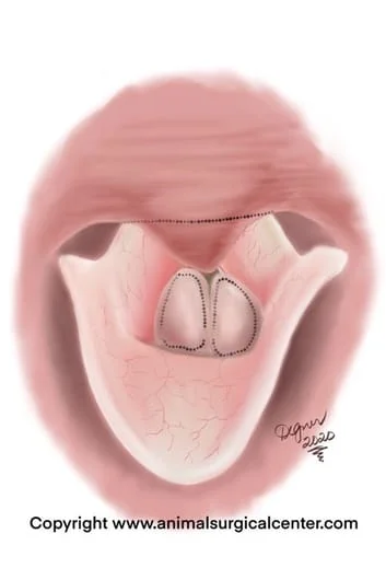
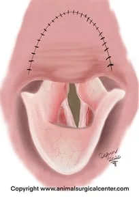
Along the side of the vocal cords is a recess or cavity called the lateral ventricles of the larynx. Brachycephalic dogs commonly have eversion of the mucosa of the lateral ventricles. These balloon-like structures occlude the opening of the larynx. See illustration right which shows a view of the larynx; Sac = everted saccule; SP=elongated soft palate. The everted saccule are removed if they are bulbous and obstruct the airway.
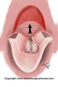
The soft palate can be shortened using a number of potential methods. It is critical to determine if the soft palate is only too long or if it is too long and thick. If the palate is thin and elongated, a simple trimming of the elongated portion of the soft palate can be done. This may be done with done with a scalpel blade/scissors, laser or Ligasure (see illustration below).
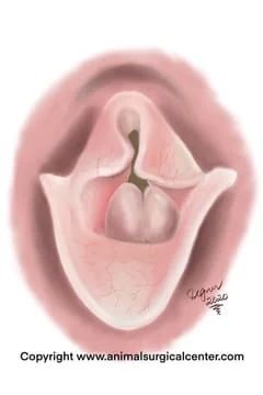
Many brachycephalic dogs not only have an elongated palate, but it is also a very thick. The only way to determine if the palate is too thick is with CT scan and for that reason, this test is routinely completed prior to surgery at ASCM. For dogs with a very thick palate, a folded palatoplasty is used. In this procedure the distal tip of the soft palate is tagged with a suture and pulled forward to determine the front extend of the incision in the soft palate. Then a section of the palate along with the muscle of the palate is resected. The tip of the soft palate is then pulled forward and the incision is sutured closed. The end result is a thin and short soft palate (see below). After the surgery, gauze sponges soaked in mannitol are applied to the tissues for about 20 minutes after surgery (while the patient is still anesthetized) to help reduce swelling.
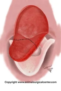
These illustrations demonstrate the folded palatoplasty as seen from the surgeon's point of view (oral view). If the saccules are large, they are also removed at that time.
In some patients that develop marked swelling of the throat after surgery, a life-saving temporary tracheostomy is needed after surgery, but this significantly increases patient morbidity and hospital stay.
Care after surgery
Most pets having upper airway surgery will stay in the hospital at least one night at ASCM to monitor for breathing difficulties. While in the hospital, intravenous fluid therapy likely will continue until the pet goes home. Oxygen therapy may be needed via a nasotracheal catheter. If severe breathing difficulties are present after surgery due to swelling and the oxygen therapy via the nasotracheal catheter is inadequate, a tracheostomy may be required. This necessitates making an incision over the neck and creating a hole in the trachea, through which a breathing tube (trach tube) is placed. This tube will need to be replaced every 6 to 8 hours and the tracheostomy site cleansed on a regular basis. This type of care should only be done in the hospital by an experienced team, as occlusion of the tube with mucus can create a life-threatening situation.
At home, medication to control pain after surgery should be administered for a few days. Antibiotics may be prescribed, but may be needed for a week or two, unless the pet has pneumonia. If prescribed by the surgeon, cortisone may be needed for only a very short period of time after surgery. Exercise should be restricted for two weeks, but leash walks are generally permitted. A follow-up examination should be done in 2 weeks after surgery to evaluate the healing process.
Results
Most immature dogs that have surgical correction of the problem usually recover uneventfully. As a dog ages with untreated brachycephalic airway syndrome, secondary changes such as laryngeal collapse may complicate the recovery and worsen the prognosis. Aspiration pneumonia will complicate the recovery and in some cases can be fatal. A successful surgery will result in much better breathing, snoring will be reduced, exercise tolerance is greatly improved, sleeping will be much better, and weight loss with an appropriate dietary restrictions can be achieved. Although surgery can be successful, it is imperative that the pet does not become obese, as this will result in breathing difficulties again. In some cases, especially those that have been untreated for a prolonged period of time, the larynx becomes weakened and the cuneiform cartilages collapse together (see right). In such cases, a permanent tracheostomy may be needed.
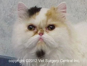
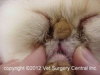
A complication that may be seen following laser surgery on the nose in very small animals, such as a cats, is permanent stricturing (narrowing) of the nares. This is caused by collateral thermal damage to neighboring tissues. Below is a cat that has stenotic nares prior to laser surgery and then the disaster that followed. Take note that the photo below right shows that the nostrils have healed closed and the nose is gone, as a result of failed laser surgery. As a warning, only select a surgeon that has good experience with this technique. Be aware that the cold scalpel blade is unlikely to have this kind of end result versus when treated with laser.
Another potential complication of soft palate surgery is resecting the soft palate too short, which results in reflux of food into the nasal cavity with subsequent chronic nasal infection.
