

Key Points
Ureteral stones obstruct the flow of urine from the kidneys to the bladder and left untreated can cause destruction of the kidney.
Surgical removal of the stone is the treatment of choice if medical therapy (intravenous fluids) not effective.
The prognosis is favorable, unless the kidney is no longer functional.

- Urine produced by each kidney passes down a tube called the ureter to a reservoir, the bladder.
- The ureters have a thick muscular wall that propel urine to the kidneys in concerted waves of contraction.
- The inner tube (lumen) of a ureter is very narrow and small stones that form in the kidneys can become lodged as they try to pass down into the bladder.
Signs of ureteral stones
- In humans, the passage of a stone down the ureter is more painful than giving birth to a child. In dogs and cats the passage of a stone down a ureter frequently goes unnoticed until the cat is ill from kidney failure.
- Clinical signs of kidney failure may include anorexia, vomiting, urination suddenly stops, lethargy, and dehydration.
- Sometimes a few stones will make their way to the bladder and then become lodged in the urethra thus the pet will not be in a lot of pain and will be able to urinate at all.
Why do stones develop?
- The most common type of stone in cats is the calcium oxalate stone. The cause of this is largely not understood, but may be a defect in the metabolism of oxalic acid or excessive spill over of calcium into the urine by the kidney. Some Schnauzers are born with a defect that predisposes them to calcium oxalate stone formation.
- Calcium oxalate stones may form if the pet has a tumor of the parathyroid gland or another type of cancer.
- Ammonium biurate stones form if the pet has a liver disease such as a portosystemic shunt.
- Urate stones form in Dalmatians due to a defect in metabolism
- Some diets encourage stone formation.
Diagnosis
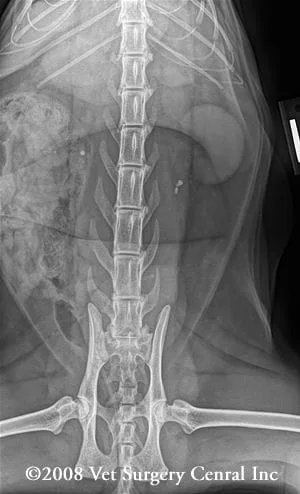
- Radiographs will confirm the presence of stones (place cursor over radiograph). The use of abdominal compression techniques while making the radiograph can make the stones more visible.
- Ultrasound of the urinary tract will show the distention of the ureter and the pelvis of the kidney. In some cases the stones will also be visible on the ultrasound.
- Nuclear scintigraphy may help to show if the kidney that is affected by the ureteral obstruction is functional, however, if the ureter is completely obstructed, the kidney will appear to be misdiagnosed as nonfunctional.
- Blood tests that are routine include a complete blood count and chemistry profile. Urine testing for bacterial infection and chemical analysis is also recommended.
Treatment
- Medical therapy includes aggressive fluid therapy. Careful monitoring of such patients is essential as they can become overloaded with fluids and the lungs will become congested to the point of causing breathing difficulties. Radiographs are made intermittently during the treatment to see if the stones will pass into the bladder. If the stones have not moving in a 12 to 24 hour period, surgery will be recommended.
- Surgical candidates for surgery include
- patients that have stones in the ureter
- dilation of the renal pelvis and ureter are noted
- the kidney on the affected side has visible tissue (not just a bag of fluid due to chronic ureteral obstruction)
- In order to have a very high success rate, the surgeon should use high magnification that is offered with an operating microscope and use very fine instruments and suture that is finer than human hair.
- Surgery entails making an incision into the affected ureter very close to the stones (typically the incision is made a bit closer to the kidney) in a dilated region of the ureter. The stones are removed with fine instruments and the ureters are flushed. If the surgeon is uncertain about the patency of the ureters distal to the obvious site of obstruction, the bladder is opened and fluids are flushed down the ureter to the bladder. The surgeon will be able to visualize the flow of fluid at the opening of the ureter in the bladder.
- After surgery, intravenous fluids will be administered to keep the patient well hydrated. Analgesics are administered to control pain. If infection is present, antibiotics may be continued for 2 to 3 weeks after surgery.
Care at home
- The incision should be checked daily for signs of infection
- Make sure that your pet is urinating well at home.
- Offer canned food instead of dry food, as this will help to prevent stone reformation
- The stones will be chemically analyzed and the surgeon may make recommendations about the type of diet that should be fed.
Prognosis
- Generally the prognosis is good for the surgery to relieve the obstruction and for healing of the ureter(s). If the patient has diseased kidneys, the kidney values on the blood work may not normalize, but frequently will stabilize and the patient will have a good quality of life.
- Stone may reform, therefore radiographs, blood and urine testing should be done every 3 months.
