

Key Points
Tumors of the oral cavity are best treated when the tumors are small
For many of the tumors aggressive is needed to obtain a cure
In spite of removing substantial sections of the jaw the appearance is still cosmetic
Reconstructive surgery can be done to restore the shape of a jaw bone using bone grafts
Prevalence of oral tumors
- 6% of all types of cancers in dogs are located in the mouth cavity
- These are the fourth most common type of cancer
Types of tumors
- Benign – don’t spread and potentially can be successfully removed
- Malignant – grow deeply into the tissues (have roots) and/or spread to the rest of the body very quickly
Oral epulis
- Do not spread to the rest of the body
- Fibromatous epulis
- usually a tumor that is located at the margin of the gums
- generally are smooth, pink and do not have an ulcerated (raw) surface
- surgical removal is generally curative
- Ossifying epulis
- tumor is smooth and nonulcerated
- can be more difficult to cure by simply cutting the mass off
- after this type of mass has been removed the bed is treated with cryosurgery (3 cycles of freezing)
- sometimes a more radical surgery is needed, which involves removal of a part of the underlying bone (partial maxillectomy, partial mandibulectomy)
- tumor may have ulcerated or smooth surface
- these tumors most commonly affect the front part of the lower jaw
- tumor originates from the ligament (periodontal ligament) that holds the tooth root in the bone
- tumor is very locally invasive and therefore is considered a type of cancer
- in order to remove the tumor a portion of the bone also needs to be removed (maxillectomy, mandibulectomy)
- with surgery these tumors have a 95% chance to be cured
- if the tumor is very small, radiation can be used
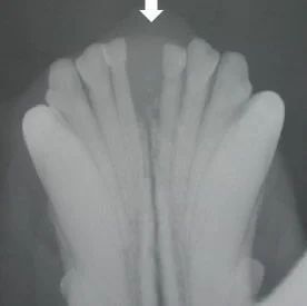
- Photo right is an x-ray of “Bella’s” front jaw – take
Locally invasive malignant tumors
- These tumors are locally invasive, that is they have roots of cancer that are deeply seated in the jaw bones
- These tumors have a lower tendency to spread to the rest of the body
- Fibrosarcoma, soft tissue sarcoma
- less than 20% spread rate to lungs
- must perform a radical surgery to attempt to remove this type of tumor
- radiation therapy is usually recommended following this type of surgery to try to kill the roots of the tumor that may be left behind
- chemotherapy may be recommended if the tumor is high grade based on biopsy results
- median survival is about 7 months with radiation alone
- survival with surgery and radiation therapy is about 1 to 2 years
- the primary goal of surgery is palliative…to buy time
- if the tumor is located in the front of the mouth there is a very good prognosis as surgery can be curative
- if the tumor is located in the back part of the mouth, tongue, or tonsils, the spread rate is very high therefore prognosis is poor
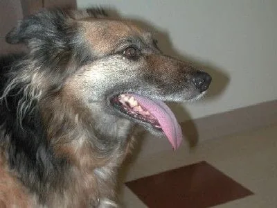
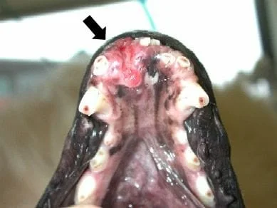
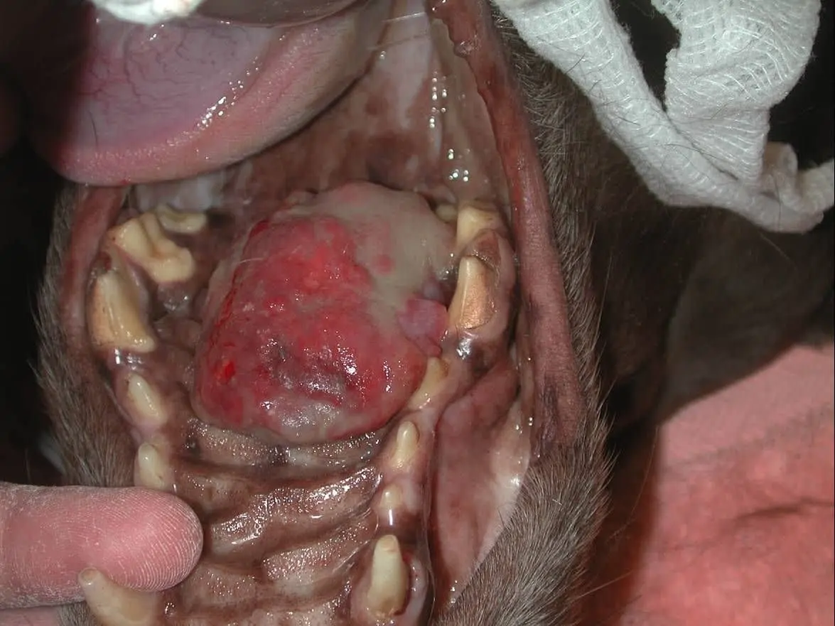
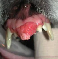
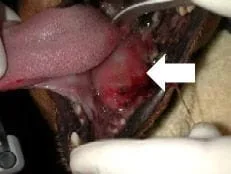
- photo right is an example of a squamous cell carcinoma on the front of the lower jaw, the excised tumor, and appearance of the patient immediately after surgery
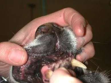
- photo below is the appearance after the front portion of the mandible has been removed. The dog had excellent function of the jaw and was cured of the disease with surgery alone.
Malignant tumors that tend to metastasize (spread)
- Melanoma
- may appear as a black mass, but some do not have the pigment (amelanotic)
- locally invasive
- at the time of diagnosis, many have already spread to local lymph nodes or the lungs
- radical local surgery is needed to remove the tumor
- if the lymph nodes are large, radiation therapy may be used as they tend to respond well to this treatment
- chemotherapy may also be recommended
- size counts with this tumor
- if the tumor is less than 2 cm in size, the median survival is 511 days
- if the tumor is greater than 2 cm, the median survival is 164 days
- the grade of the tumor on biopsy also dictates prognosis
- the stage, or evidence of spread of tumor to the rest of the body is important
- stage 1 (tumor isolated the jaw alone) – as a greater than 2 year survival
- stage 2 (tumor spread to local lymph nodes) and stage 3 (tumor in nodes and lungs) – have a poor prognosis
- if this type of tumor is located on the upper jaw (maxilla) the prognosis is generally poor with mean survival of about 5 months due to spread of tumor to the lungs and other parts of the body (in this set of cases only surgery was performed); our oncologist has seen a mean survival of about 1 year with surgery and chemotherapy
- the photo right is a case of osteosarcoma of the roof of the mouth (hard palate)
- if this tumor is located on the lower jaw (mandible) the prognosis is much better and surgery can be potentially curative; one report showed a 71% survival rate at 1 year after surgery (this means that nearly 3/4 of the dogs were still alive at 1 year!)
Diagnostic tests
- Complete blood cell count, Chemistry profile, Urinalysis are performed to evaluate the overall health of the patient
- Chest radiographs (x-rays) are used to help see if the tumor has spread to the lungs
- Oral radiographs (x-rays) are taken of the tumor site to see how invasive the tumor is in the bone
- Biopsy of the tumor – a small piece of the tumor is sent for analysis by a pathologist to determine the type of tumor that is present as this will give an idea on the prognosis
- If any lymph nodes are enlarged, a fine needle biopsy is done to help determine if there is spread to the nodes
- After surgery has been completed, the entire tissue specimen is sent to the lab to check the margins to ensure that all of the cancer has been removed
- Maxillectomy – removal of the upper jaw
- Mandibulectomy – removal of the lower jaw
- In some cases the entire jaw bone is removed and in other cases in which the tumor is smaller, only a portion of the jaw is removed
- In spite of removing relative large portions of a jaw bone, the cosmetics are still very good
- Dr. Degner has the capability to rebuild a jaw bone using microvascular surgery and transplantation of a bone graft from the shin bone or other bones.
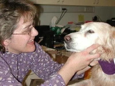
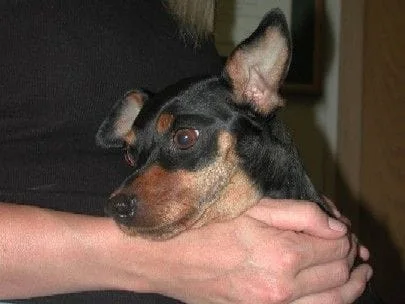
- In the photo right (put cursor on image to see removed part), the front part of the upper jaw was removed to treat an acanthomatous epulis; take note of the very good cosmetic appearance of the “Duchess” three years after surgery
Aftercare
- Feed soft food for about 3 weeks or until your veterinarian indicates that the incisions have healed well
- No chew toys or raw hides for 3 weeks
- Limit activity – no rough play until your veterinarian indicates that it is safe to return to normal activity
- Hot pack the face three times daily 10 minutes each time until the swelling resolves
- Flush mouth with water or antiseptic such as Nolvadent after eating
- If malignant tumor was removed chest radiographs should be taken every 3 months
- If chemotherapy or radiation is recommended, schedule an appointment with an oncologist

