

Key Points
If the growth plate of the radius bone is damaged while a dog is growing, there is a significant risk that the patient may develop a shortened radius bone which ultimately causes the elbow joint have an imperfect fit.
Treatment of this condition usually involves removing a segment of the ulna bone, just below the elbow.
Surgery usually will dramatically improve the lameness, yet heavy exercise in these patients may cause some stiffness or lameness.
Pertinent anatomy
- The elbow joint is formed by the the ends of the humerus bone, radius bone and the ulna. It is critical that each bone fits perfectly within the elbow joint so that abnormal wear and tear on the joint does not occur. In addtion, abnormal stresses may be placed on coronoid process, may cause this part of the bone to break off (called a fragmented coronoid process).
Pathology of a shortened radius bone
- The long bones of the body have a growth plate located at each end. The longitudinal growth of the bone occurs at the growth plate. If the growth plate is damaged while a dog is growing (major growth in large breed dog takes place from 4 to 8 months of age), then the affected bone may be shorter than normal.
- If the growth plate of the radius (either the top or bottom one) is damaged the radius bone becomes shortened. The result frequently is two fold:
- the elbow gets pulled out of alignment and more weight is then put on the medial and lateral coronoid processes, thus these processes may break off
- the carpus (wrist) may develop abnormal twisting
Signs of a shortened radius bone
- Most dogs are under one year of age when this problem is first dagnosed
- Breeds commonly affected are Bernese Mountain Dogs and less commonly Retrievers and Mastiffs
- Affected dogs are commonly lame on the affected forelimb. These dogs may externally rotate the forelimb in order to relieve elbow pain. In other cases, the affected limb may have a true rotational abnormality due to twisting of the bones from abnormal growth.
- The elbow may be swollen and painful on direct palpation.
Diagnosis
- Frequently a shortened radius can be seen on plain radiographs (x-rays) of the affected forelimb (see below)
- CT scan may also be used to help diagnose secondary issues such as a fragmented coroinoid process within the elbow joint
Treatment
- The simplest treatment for this condition is to remove a segment of ulna bone, just below the elbow joint. The amount of bone that is removed is inversely proportional to the dogs age. In other words, if the dog is young (ie about 5 months old) then a much larger segment of bone should be removed from the ulna bone versus a dog that is no longer growing (about 10 to 12 months old). I prefer to place a pin in the ulna bone for alignment and also pull the cut ends of the ulna bone together with a heavy nylon suture so that that elbow joint will assume a better fit immediately after surgery. The space between the cut ends of the ulna bone eventually heal together, but this process may take about 3 months.
- A more complex form of treatment is to lengthen the radius bone, but this should be reserved for dogs that have stopped growing.
- Regardless of the type of treatment used to make the elbow joint fit better, arthroscopic examination of the joint is recommended at the time of surgery to rule out the possiblity of a fragmented coronoid process. If this is the case, the loose fragment of bone is removed from the joint at that time.
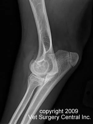
Preop radiograph showing a shortened radius and a 6 mm of radiohumeral incongruity
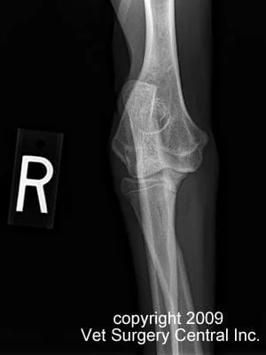
Preop radiograph showing a shortened radius with the coronoid process riding high
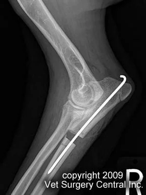
Immediate postop radiogaph showing dramatic improvment of the congruity of the elbow joint after a proximal partial ulnar osteoctomy. In this case 1.5 cm of bone was removed and a pin placed.
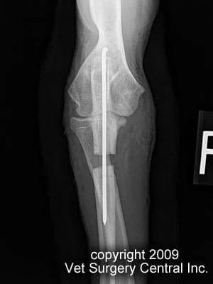
Immediate postop radiograph showing dramatic improvement of the congruity of the elbow joint. Take note of the poximal ulnar ostectomy site and the which aligns the ulna. The step in the elbow has been eliminated.
Prognosis
- A report of 11 dogs that were treated for shortened radius bone syndrome for the most part were significantly improved. Lameness associated with heavy activity was noted in most dogs.
References
- Vandewater A and Olmstead ML. Premature closure of the distal radial physis in the dog: a review of eleven cases. Vet Surg 1983;12 (1):7-12
- Cook CR, Cook JL. Diagnostic imaging of canine elbow dysplasia: a review.Vet Surg. 2009 Feb;38(2):144-53
- Samoy Y, Van Ryssen B, Gielen I, Walschot N, van Bree H. Review of the literature: elbow incongruity in the dog. Vet Comp Orthop Traumatol. 2006;19(1):1-8. Review.
- Preston CA, Schulz KS, Taylor KT, Kass PH, Hagan CE, Stover SM. In vitro experimental study of the effect of radial shortening and ulnar ostectomy on contact patterns in the elbow joint of dogs. Am J Vet Res. 2001 Oct;62(10):1548-56
- Ubbink GJ, Hazewinkel HA, van de Broek J, Rothuizen J. Familial clustering and risk analysis for fragmented coronoid process and elbow joint incongruity in Bernese Mountain Dogs in The Netherlands.Am J Vet Res. 1999 Sep;60(9):1082-7.
- Boulay JP. Fragmented medial coronoid process of the ulna in the dog. Vet Clin North Am Small Anim Pract. 1998 Jan;28(1):51-74.
