

Key Points
Mild to moderate lameness can be associated with calcium deposits in the supraspinatus tendon.
Treatment involves removal of the calcium deposits from the tendon.
New treatment!!!! Stem cell injection in the supraspinatus tendon lesions is highly successful – may be better than surgery!
Prognosis generally is good.
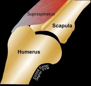
Anatomy
- The shoulder is supported by tendons and ligaments.
- The supraspinatus tendon extends the shoulder joint.
Damage to the infraspinatus tendon
- Injury to the supraspinatus tendon frequently results in inflammation of the torn part of the tendon
- As a result of the inflammation, calcium deposits form in the tendon. The result is a chronic source of pain for the patient
Signs
- Lameness frequently is associated with exercise
- The degree of lameness frequently is mild to moderate
- Sometimes all that is noted is that the dog will not swim in a straight line, rather tends to turn toward the affected limb.
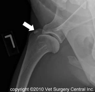
Diagnosis
- The diagnosis of supraspinatus tendinopathy is made by identifying calcium deposits in the supraspinatus tendon on radiographs (x-rays)
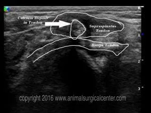
- Some dogs will not have obvious calcification on radiographs, therefore ultrasound should always be considered as a part of the work-up for these patients. The ultrasound images below are of a dog that had only a hint of mineralization of the supraspinatus tendon, but marked changes are seen within the tendon
- Some dogs will have the calcium deposit in the supraspinatus tendon and are asymptomatic for this problem. Nearly all dogs that have a clinical lameness due to the calcium deposit do not seem to be overtly painful when the tendon is palpated or stretched by flexing the shoulder maximally. For this reason the veterinarian must make sure that there is no other problem that is causing the lameness of the limb (fragmented coronoid process, osteochondritis dissecans, biceps tendon tear, shoulder ligament tear, teres muscle tear etc) before recommending surgery.
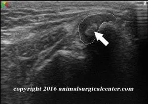
Transverse image of supraspinatus with calcification
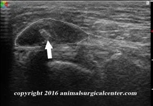
Sagittal view of supraspinatus tendon with calcification
Treatment
- Surgery
- An incision is made over the region of the affected tendon. An incision is made to separate the fibers of the supraspinatus tendon and the calcified material is removed.
- The clinical signs of supraspinatus tendinopathy resolve because
- the irritating calcium deposit has been removed
- the tendon is opened up to allow a blood vessels to grow into the injured area and thus promote healing
- Stem cell with platelet-rich plasma treatment
- Stem cells from bone marrow or fat can be injected with PRP into the diseased tendon.
- Prevention of tendinopathy – dogs need to have their muscles and tendons stretched prior to heavy exercise. Stretching the supraspinatus involves flexing the shoulder maximally.
Prognosis
- Most dogs that have surgery for this condition respond well.
- Stem cell treatment has been shown to be 97% effective AND THIS MAY BE MORE SUCCESSFUL THAN SURGERY.
- Lameness usually resolves within 2 months after surgery.
- Recurrence of the problem may occur if the pet reinjures the tendon again, but in general this is not common.
