The superficial digital tendon runs along the back side of the Achilles’ heel.
After sustaining a tear to the tissues that hold this tendon in place, the tendon can dislocated of to the side of the heel bone which frequently will produce a popping sound. In addition, the patient will be lame on the affected limb.
Surgery is recommended to treat this condition
The prognosis is very favorable for these patients.
Anatomy
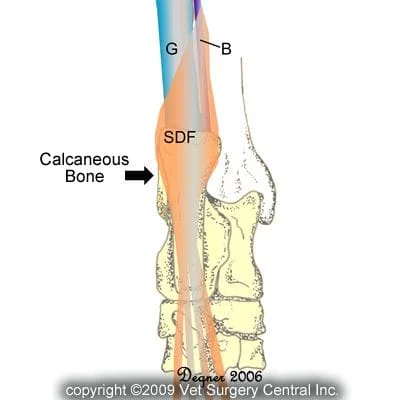
The Achilles’ tendon consists of a series of tendons that attach onto the calcaneus bone (Achilles’ heel) and the superficial digital flexor tendon (SDF) tendon that passes directly over the back side of the Achilles’ tendon and then attaches onto the bones of the toes in the hind paw.
See illustration right which demonstrates the anatomy of the tendons that extend over the back side of the hock (key: SDF = superficial digital flexor tendon; G = gastrocnemius tendon; B = biceps femoris tendon). Take note that the biceps femoris and gastrocnemius tendons attach directly onto the calcaneus bone, whereas the SDF tendon runs over the calcaneous bone. The SDF tendon tendon is held in place by tough tissue along both sides of the tendon called retinaculum.
Pathology
Trauma is the most common inciting cause for a tear of the retinaculum which holds the SDF tendon in place.
In some cases, the shape of the end of the calcaneous bone may be abnormal which causes the tendon to naturally dislocate without trauma. This problem is seen most commonly in Shelties.
When the tendon pops off the side of the bone, pain results and the dog may cry out or become acutely nonweight-bearing
Signs of SDF tendon luxation
Sudden onset of lameness
Intermittent severe lameness with interposed periods of mild lameness
An intermittent popping sound may be heard each time the tendon dislocates
Swelling over the back point of the hock (Achilles’ heel) is common
Diagnosis
The diagnosis of the SDF tendon luxation is made on physical examination in which the tendon can be popped back and forth over the Achilles’ heel (see below left)
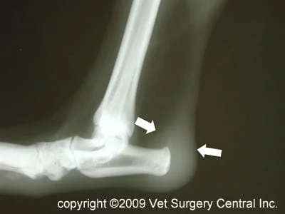
Radiographs may be recommended to rule out fractures or other problems with the bones. Usually the only finding is swelling over the Achilles’s tendon (see below right)
Preop blood work is generally done to ensure that your pet is in good internal health for anesthesia
Treatment
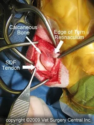
An incision is made over the site of the torn retinaculum and the tear is identified (photo below left)
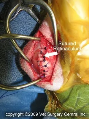
The torn retinaculum is sutured (photo below right)
Below is a video clip that shows the tendon luxating during surgery and suturing of the tendon.
Care at home
Exercise must be limited for 2 months.
A soft padded bandage is applied to the hind limb for a period of 2 weeks. A splint may also be used to support the repair during the healing period.
Pain medications and a nonsteroidal anti-inflammatory likely will be prescribed.
Prognosis
Most patients that have a simple traumatic tear of the retinaculum respond very well to the surgery and regain normal function of the limb.
Dogs that have an abnormally shaped tip of the calcaneus bone may be more prone to recurrence of the luxation.