

Key Points
Skin grafts are an excellent modality used to reconstruct wound located on the limbs and other areas of the body
With ideal wound conditions, 95 to 100% survival of the skin graft is expected
After healing takes place, the skin graft should provide a durable covering over the wound
Definition of a skin graft
- A skin graft is a piece of skin that has been totally removed from the body and placed on a wound. Blood vessels in the wound bed will quickly grow into the underside of the skin graft, thus bringing it back to life. If the blood vessels cannot grow quickly enough into the the underside of the skin graft, the graft will die. For this reason the wound must be adequate to nutritionally support the graft and the surgeon must very carefully prepare the skin graft so that vessels will be able to grow into the graft.
Indications for skin grafts
- Skin grafts are used for wounds that are caused by
- traumatic accidents
- oncological surgery (tumor removal)
- thermal burns
- chemical burns
- vesicant burns – injected medications such as chemotherapeutic medications or some injectable anesthetics
- The procedure has the advantage of requiring only one surgery once the wound bed is adequately prepared for grafting.
Contraindications for skin grafts
- Skin grafts frequently do not “take” (die) in the following cases
- radiation has been administered to the area prior to surgery
- over bone
- over tendons and ligaments
- poorly vascular wound beds
- high motion locations that cannot be immobilized
- infection in the wound
Types of skin grafts that are used to repair wounds
- Full-thickness skin grafts are most commonly used in dogs and cats. This involves removing the a piece of skin and removing the fat from the underside of the skin. The donor site must have enough surrounding loose skin so that the incision can be closed. The survival (“take”) of a full-thickness skin graft is the same as a partial-thickness skin graft.
- Partial-thickness skin grafts skin grafts involve shaving a very thin layer of skin off the donor site. No hair will grow from this skin graft because the hair follicles are in the deeper layers of the skin. The donor site will heal on its own and does not involve closure of an incision. This type of graft donor site can be more painful, as the raw donor site will have many exposed nerve endings that need to heal over with time. The indication for partial thickness skin grafting is for cases in which a dog has massive skin loss (especially a burn victim) and there is limited normal skin that is available to be used for skin grafting.
Phases of healing of a skin graft
- Imbibition: the small blood vessels on the under side of the skin graft soak up fluid (serum) from the wound bed,which provides oxygen and nutrients during the first 2 days after surgery.
- Inosculation: to “inosculate” means to “kiss”. Small blood vessels (capillaries) in the wound bed meet up (kiss) and connect with cut vessels of the skin graft. At this time blood begins to circulate in the skin graft and provide it nutrition. Inosculation typically occurs within 48 hours after surgery, depending on the condition of the wound bed.
- Revascularization: small capillary blood vessels bud and grow into the capillary vessels of the skin graft like a snake would slide down a tunnel, thus providing a great source of blood to the skin graft. Revascularization of the graft takes place from day 4 to 7 after surgery.
Wound preparation and skin grafting procedure
- Skin grafting requires a fastidious bed to provide an excellent “take” of the skin
- A wound bed that is ideal for a skin graft is granulation tissue (pink red healing tissue), muscle or fascia over a muscle.
- If the wound bed does not have an ideal surface for skin grafting, the procedure will be delayed until a healthy granulation tissue bed has developed. This may take about 7 to 10 days. For example if a skin tumor has been removed from a limb, the wound can be covered with a bandage and the grafting delayed until granulation tissue is present. The bandage will need to be replaced daily until skin grafting is performed. Similarly, if a dog sustains a wound due to trauma, the grafting will be delayed until healthy granulation tissue is present.
- If the granulation tissue is excessive, it will be trimmed down to the surface of the edges of the skin and the grafting procedure is delayed for four days.
- On the day of the procedure your pet will be anesthetized and a site will be selected from which a skin graft will be collected. This site typically is the side of the abdomen or chest region, as there is abundant skin that can be safely removed without ill effects to your pet.
- As the skin graft is being elevated the fat will be carefully cut off the under side of the skin.
- Small holes will be made in the skin graft to allow fluid to escape from beneath the flap. The accumulation of fluid under the graft will prevent separate the skin from the granulation tissue and prevent the ingrowth of blood vessels into the graft.
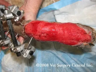
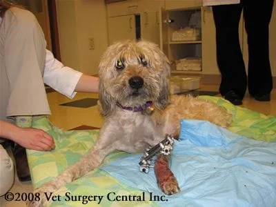
- The skin graft is stapled or sutured over the wound (see photo left) and the limb is bandaged.
- After surgery, it is imperative that the grafted are is bandaged and supported with a splint to prevent motion at the graft site.
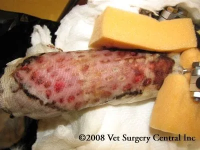
- The first bandage is typically kept in place for about four to five days after surgery. Care is taken when changing the bandage so that the skin graft is not pulled off the wound bed with bandage material that may be adhered to the skin.
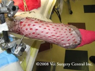
- In the photo right (4 days after surgery), the dressing layer that is stuck on the graft is not removed, rather triple antibiotic ointment is applied over this layer and the limb is rebandaged.
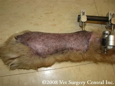
- By seven days after surgery, the graft should be well secured to the granulation tissue bed and all layers of the bandage can be removed.
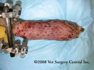
- The skin graft in the photo right is seen 18 days after surgery. Approximately 99% of the graft has survived and the dog is doing very well.
- Skin grafts provide a thin covering over distal extremities, therefore are very cosmetic.
- Hair growth on the skin graft may be sparse, which may be related damage to the hair follicles during collection of the skin graft. The hair usually can be oriented in the appropriate direction to match that of the native coat of the limb.
Care at home
- Limit activity for 3 weeks after surgery
- Keep the bandage dry
- Check the toes for swelling and coldness if the graft site is on a limb (while the bandage is on)
- Return to your veterinarian at the scheduled times for bandage changes
- After the bandage is no longer needed, cover the graft site with a sock (if the graft site is on a limb) for a period of 3 to 4 weeks to prevent your pet from licking and chewing at the site. If needed, an Elizabethan collar may be recommended to prevent self-mutilation
Prognosis
- With an ideal wound bed, about 95 to 100% “take” of the skin graft is expected.
- Survival of the skin graft is dependant on restricting your pets activity, especially during the first week
- Skin grafts frequently do not grow hair, but provide a durable covering over the wound (see photo right of a skin graft 7 weeks after surgery).
References
- Guille AE, Tseng LW, Orsher RJ. Use of vacuum-assisted closure for management of a large skin wound in a cat. J Am Vet Med Assoc 2007; 230-1669-1673.
- Aragon CL, Harvey SE, Allen SW, McCrackin-Stevenson MA. Partial thickenss skin grafting for large thermal skin wounds in dogs. Comp Contin Edu Pract Vet 2004; 200-215.
- Carson-Dunkerley SA, Hanson RR. Equine skin grafting: principles and field application. Comp for Cont Education for Vet; 1997.
- Swaim SF: Skin Grafts in Swaim SF (ed): Surgery of Traumatized Skin: Management and Reconstruction in the Dog and Cat. Philadelphia, WB Saunders Co 1980, pp 423-476.
- Lees MJ, Fretz Pb, Bailey JV, Jacobs KA: Principles of grafting. Compend Contin Educ Pract Vet 1989; 11:954-961.
- Converse JM, Smahel J, Ballantyne Dl, et al: Inosculation of vessels of skin graft and host bed: A fortuitous encounter. Br J Plast Surg 1983; 29:274-282.
- Ross GE. Clinical Canine skin grafting. J Am vet Med Assoc 1986; 153:1759-1765.
- Bauer MS, Pope ER: The effects of skin graft thickness on graft viability and change in original graft area in dgos. Vet surg 1986;15(4):321-324.
- Pope ER: Effect of skin grat prparation and graft viability on the secondary contraction of full-thickness skin grafts in dogs. Am J Vet Res 1985; 46(12)2530-2535.
- McKever PJ, Braden TDD: Comparison of full- and partial-thickness autogenous skin transplantation in dogs: A pilot study J Am Vet Res 1978; 39(10):1706-1709.
- Probst CW, Peyton LC, Binham Hg, et al. Split-thickness skin grafting in the dog. J Am Anim Hosp Assoc 1983;19:555-568.
