

Key Points
Salivary mucocele is an accumulation of saliva outside of the salivary gland that is due to rupture of a salivary duct.
The swelling within the tissues due to the mucocele can be under the tongue, in the back of the throat, around the eye or just behind the lower jaw (on the under side of the neck).
Surgery is the treatment of choice and results in an excellent prognosis
Anatomy
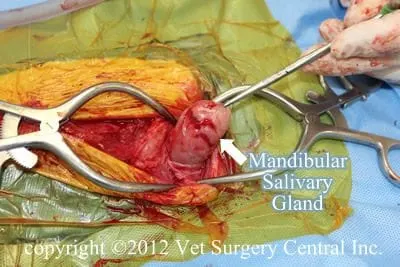
Dogs have numerous salivary glands located in a variety of areas near the mouth cavity. The mandibular salivary glands, located just behind the lower jawbone, are connected to the sublingual salivary glands. These glands have ducts which open on the underside of the tongue. The zygomatic salivary glands are located in the eye sockets. The parotid salivary gland is located over the ear canal and the side of the cheek. These later two glands open on the inside of the upper lip.
Salivary mucocele
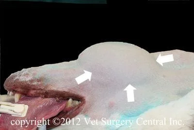
A cervical mucocele is due to rupture of the mandibular/sublingual salivary duct complex.
A ranula is a collection of saliva beneath the tongue and is due to rupture of the sublingual salivary duct.
A pharyngeal mucocele is a collection of saliva in the back of the throat and is due to rupture o the mandibular/sublingual ducts.
A zygomatic mucocele is a collection of saliva around the the eye and due to rupture of the zygomatic salivary gland 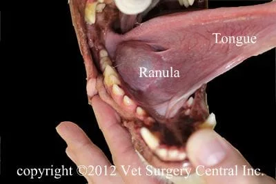
Signs
Breeds predisposed to this condition include Dachshunds, Poodles and many other breeds; cats infrequently are effected by this condition. This condition can occur at any age. The typical signs of a salivary mucocele include a soft, nonpainful swelling located on the under side of the neck, under the tongue, in the back of the throat or around the eye. A large ranula, may get traumatized by the teeth and bleed. Pharyngeal mucoceles may cause swallowing or breathing difficulties. The cervical (neck) mucoceles usually have no signs other than a swelling located just behind the lower jaw on the under side of the neck. Mucoceles are typically nonpainful, unless a secondary bacterial infection is present.
Diagnosis
Fine needle aspiration reveals a thick ropy fluid that is commonly blood-tinged. Examination of the fluid under the microscope frequently shows chronic inflammation (mononuclear cells) and red blood cells. The diagnosis is confirmed with exploratory surgery and finding the ruptured duct. In preparation for surgery, preoperative blood work including a complete blood count, chemistry profile, and urine testing are recommended to ensure that your pet is healthy and can safely undergo anesthesia.
Preparation for surgery
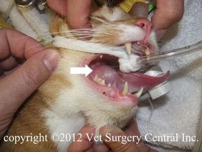
The pet should be fasted prior to surgery, as instructed by the surgical team. Water is usually permitted up to the time of admission to the hospital. An antacid such as Pepcid AC may be prescribed and should be administered by 6 AM on the day of surgery; this treatment will help reduce the risk of esophagitis (heartburn) in the postop period. The surgical team should be informed of any medications that your pet is currently receiving. The pet should not receive any aspirin within 1 week of surgery, as this medication will thin the blood and increase the risk of bleeding. Just prior to surgery, your pet will receive a sedative, have an intravenous catheter placed for the administration of intravenous fluids and intravenous medications, be induced under general anesthesia with medication(s), and have a breathing tube (endotracheal tube) placed to allow delivery of oxygen and gaseous anesthesia. The surgical site will be clipped and cleansed with an anti-septic solution in preparation for surgery. While under general anesthesia, the pet’s breathing will be assisted with a ventilator and vital parameters such as heart rate, respiratory rate, core body temperature, blood pressure, oxygenation of the blood (pulse oximetry), exhaled carbon dioxide (capnography), and heart rhythm (EKG) will be monitored to ensure the pet’s well being. Pain will be controlled both during and after surgery with analgesics (pain-controlling medication). Please note that each surgical and anesthesia team may elect to chose a different, but effective analgesia protocol.
Surgery
Aftercare
Following surgery, the patient will receive pain-relieving medication to ensure a comfortable recovery. A combination of nonsteroidal anti-inflammatory, local anesthetics and narcotics are used to control pain. Intravenous fluid therapy is administered to ensure that your companion will remain well hydrated during and after surgery. Most patients can go home on the day of surgery. If an invasive and more painful surgery was performed, the pet may need to be hospitalized overnight to control pain. A drain tube may be placed in the wound and should be removed once the fluid production is minimal (10 ml/24 hours); if the drain has been in place for 10 days and there still is drainage, the drain removed, as it could be inciting fluid production (foreign body reaction). At home, the incision should be checked for signs of infection. Your pet should not scratch the incision, as this could open the incision or cause infection. If necessary, an Elizabethan collar can be placed on your companion to protect the surgical site. Exercise should be restricted for about 3 weeks after surgery.
Prognosis
The prognosis following surgery is excellent with recurrence of the mucocele being very uncommon.
Complications
Complications of the procedure may include infection, recurrence of the mucocele (not all of the important tissue was removed), damage to the lingual nerve which will cause a loss of sensation to one side of the tongue (and potential chewing on tongue if the dog cannot feel it), damage to the hypoglossal nerve which will cause deviation of the tongue to the unoperated side, and profound bleeding if an important artery has been cut. Breathing difficulty may also be seen if excessive postop swelling or extensive bleeding into the surgical site occurs.
