

Key Points
If your dog or cat is showing signs of “heat” or estrus after she has been spayed, ovarian remnant syndrome is very likely the underlying cause
Clinical signs are usually all that is needed to diagnose the problem, however, vaginal cytology or hormone assays can confirm the diagnosis
The most accurate testing to confirm a diagnosis of ovarian remnant syndrome is simultaneous anti-Muellerian and progesterone hormone testing.
Surgical removal of the remnant will resolve the problem
Anatomy
The female reproductive tract of dogs and cats consists of two ovaries, uterus, and vagina. The uterus is has a short body and two long horns. The ovaries are located at the end of each uterine horn.
What is ovarian remnant syndrome?
This condition results signs of heat cycle (estrus) from elevated estrogen levels after being neutered (spayed). Failure to remove all ovarian tissue (part or entire ovary) at the time of the original spaying procedure is the most common cause for this syndrome. Some animals will have ectopic ovarian tissue, meaning that the pet was born with an extra piece of ovarian tissue that was not in its normal location rather located in the broad ligament of the uterus or another location. Hence, when the spaying procedure is performed, this piece of tissue may be left in the abdomen. Off all complications of ovariohysterectomy, 17% of these are ovarian remnants.
Signs
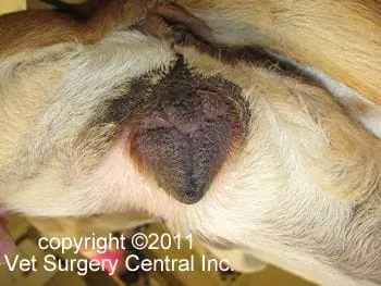
Animals that have ovarian remnant syndrome will go through a heat cycle. The average time after spaying until a heat cycle is seen is 15.5 months (range of 3 months to 5 years). Signs of a heat cycle include a swollen vulva (below left), bloody discharge from the vulva, and attraction to male dogs. Following the “heat cycle”, the nipples and mammary glands may become enlarged due to a pseudopregnancy (photo below right).

Diagnosis
Signs of a heat cycle in a spayed dog is usually consistent with ovarian remnant syndrome. If the pet receives estrogen, signs of a heat cycle also could be present; this could even occur if the owner is using transdermal estrogen cream on herself and this area of her body is contacting her pet’s skin.
Vaginal cytology (examination of cells from the vagina under the microscope) can be useful to confirm that the pet is going through a head cycle.
Sex hormone assays can be run on a blood sample collected from the affected animal to see if she has functional ovarian tissue in her body. Progesterone levels will be high for about 2 months after the signs of heat subside. The best time to submit a sample of blood for progesterone blood level is 2 weeks after the signs of the heat cycle have abated.
Luteinizing hormone blood level is also a very good test confirm that an animal has functional ovarian tissue. In the intact female (nonspayed), there is negative feed-back on the ovary from the pituitary gland (likely from gonadotropin releasing hormone). Thus, a low LH level is found in the intact female. Only for a 24 hour period during ovulation (release of eggs from the ovary) will the LH level be high. In a female that has no ovarian tissue (spayed and no ovarian remnant), the negative feed-back from the ovarian tissue to the pituitary is gone, thus the LH level is always high. Therefore, if ovarian remnant syndrome is suspected, but the LH level is high, draw blood again and test the LH level two days later.
Anti-Muellerian hormone test can accurately identify pets that have ovarian remnants. Anti-Muellerian hormone is produced solely from ovarian tissue in females. However, if the female is in the luteal phase of the heat cycle, the hormone is not produced and the ovary switches to produce progesterone. As a result, combining anti-muellerian hormone and progesterone hormone assays will greatly reduce false negatives when based on anti-muellerian test alone. Currently, the combination of anti-muellerian and progesterone hormone assays are believed to be the most accurate method of identifying ovarian remnant syndrome in pets.
After determining that there truly is an ovarian remnant, the next step is to identify where it is located. Our best test for this is CT scan. Sometimes the ovarian remnant is not located where it typically would be expected: just behind the kidneys. For this reason CT scan is routinely performed on patients having ovarian remnant syndrome in our practice.
Preparation for surgery
Make sure that your pet is fasted, as instructed by your pet’s surgical team. Water is usually permitted up to the time of admission to the hospital. Your pet’s surgeon may prescribe an antacid such as Pepcid AC, which should be administered by 6 AM on the day of surgery; this treatment will help reduce the risk of esophagitis (heartburn) in the postop period. Inform the surgical team of any medications that your pet is currently receiving. Your pet should not receive any aspirin within 1 week of surgery, as this medication will thin the blood and increase the risk of bleeding. Prior to surgery, your pet will receive a sedative, have an intravenous catheter placed for the administration of intravenous fluids and intravenous medications, be induced under general anesthesia with medication(s), and have a breathing tube (endotracheal tube) placed to allow delivery of oxygen and gaseous anesthesia. The surgical site will be clipped and cleansed with an anti-septic solution in preparation for surgery. While under general anesthesia, your pet’s breathing will be assisted with a ventilator and vital parameters such as heart rate, respiratory rate, core body temperature, blood pressure, oxygenation of the blood (pulse oximetry), exhaled carbon dioxide (capnography), and heart rhythm (EKG) will be monitored to ensure your companion’s well being. Pain will be controlled both during and after surgery with analgesics (pain-controlling medication).
Treatment
Laparoscopic examination of the abdomen or abdominal exploratory with a traditional large abdominal incision is needed to locate and remove the ovarian remnant. See ovarian remnant in the abdomen (below left) as seen with laparoscopy. The photo below right is the removed ovarian remnant in this patient.


Reference
Lofsted RM, Vanleeuwen JA. Evaluation of a commercially available luteinizing hormone test for its ability to distinguish between vasectomies and sexually intact bitches. J Am Vet Med Assoc. 2002 may 1:220 (9):131-135.
Place, N. J., Cheraskin, J.-L., & Hansen, B. S. Evaluation of combined assessments of serum anti-Müllerian hormone and progesterone concentrations for the diagnosis of ovarian remnant syndrome in dogs. Journal of the American Veterinary Medical Association, 2019; 254(9), 1067–1072.
https://doi.org/10.2460/javma.254.9.1067
