Key Points
Myelogram involves injecting dye into the sac around the spinal cord and making x-rays of the spine; if a mass or slipped disc is present, the surgeon will see a disruption in the dye column
CT scan is a special type of x-ray that provides a cross-sectional image of the spine; this test is good for diagnosing a slipped disc that is calcified
MRI is a noninvasive tool that is very useful to diagnose many problems of the spine
Myelogram
- The word myelogram is derived from 2 root words: myelo (spinal cord) and gram (picture).
- The spinal cord cannot be seen with plain x-rays of the spine.
- A myelogram involves injecting contrast media (dye) into the bag (called the dural sac..subarachnoid space) that surrounds the spinal cord.
- After the injection, the outline of the spinal cord becomes very distinct, thus allowing the neurologist and surgeon to see if there are any masses compressing the cord or if the cord is swollen.
- This test is an anatomical test, therefore it does not give us an indication of the function of the spinal cord.
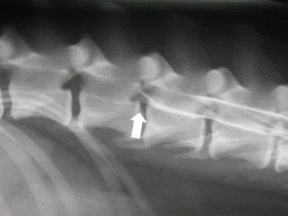
- Below is a myelogram of a dog’s thoracolumbar spine. Take note of the two white lines in the middle of the spine which is dye in the subarachnoid space. The dye which outlines the spinal cord abruptly stops due to a mass of herniated disc material. At the site of the herniated disc material the bottom dye line deviates upward because the mass of disc material is beneath the spinal cord.
- Prior to the myelogram, your pet will undergo a complete neurologic examination, blood work, and in some cases radiographs of the chest (to look for cancer of the lungs).
- Because a myelogram involves an injection of contrast media near the spinal cord, it is performed with your pet under general anesthesia. This will allow proper positioning of the pet for the x-rays, so that the best study can be obtained. In addition, your pet will not experience any discomfort from the injection
Myelogram Procedure
- An intravenous catheter will be placed and your pet then anesthetized. Very safe gas anesthetics are used during the procedure. Intravenous fluids are administered to all myelogram patients during anesthesia and recovery. Once your pet is anesthetized, a series of spinal x-rays will be taken. They will be examined for any grossly evident problems.
- The next step is the injection of contrast media. An area over the lower back or sometimes the upper neck will be shaved and cleansed with antiseptics. A spinal needle is inserted between two vertebrae (spinal bones) until spinal fluid starts to drip out. Whenever possible, a sample of spinal fluid is collected for later analysis if indicated. The contrast media is then slowly injected around the spinal cord. The contrast agent will flow throughout the spinal fluid until it outlines most or all of the spinal cord. A series of spinal x-rays is then taken. The contrast media will outline the spinal cord and indicate any area of spinal cord swelling, compression or other abnormalities. Most of the patients that have a myelogram will also have a CT scan of the area of interest to better define the problem. Since we have a CT machine in our hospital this scan is done immediately following the myelogram.
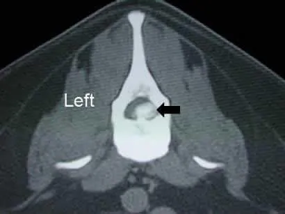
- Below is a CT scan of the same patient as above at the level of the herniated disc. This is a cross section through the spine; the oval hole in the spine is the spinal canal and the large amount of white material in the canal is herniated disc material; the smaller black crescent in the spinal canal is the spinal cord that is compressed to the top and left.
Indications for Myelography
- Myelograms are most often used to diagnose a ruptured or herniated disc. While a patient may be presented with fairly obvious signs of a disc rupture, the myelogram is still needed to differentiate a ruptured disc from other conditions.
- They are also used to diagnose the degree of spinal cord damage with a spinal fracture, tumors of the spinal cord and nerve roots, and tumors of the spine itself. In some situations a tumor around the spinal cord will have a similar appearance to that of a ruptured disc.
- The myelogram also demonstrates the precise location of the problem. This is very important so that the surgeon knows the exact location to operate the spine.
Potential complications
- As with any general anesthesia, complications may arise. Even though rare, anesthetic death can occur. With the use of modern anesthetic protocols and monitoring devices, the risk of problems with anesthesia is minimal.
- Temporary loss of breathing due to irritation of the brain stem can be caused by the contrast media. This is rare and usually transient as the contrast media is excreted from the body fairly rapidly.
- Allergic reaction (rare occurrence) to the contrast media can cause death.
- Worsening of the function of the spinal cord may occur due to inflammation or damage to the spinal cord from the injection. If this occurs, it is usually transient.
- Seizures can occur if the pet is sensitive to the contrast media. If this occurs during recovery from anesthesia, medication is used to control this. Seizure activity is almost always very transient and not fatal.
- Sometimes a nondiagnostic (non-useful) myelogram will result if the contrast media is injected outside of the sac that surrounds the spinal cord. The study may need to be repeated the next day, once the dye is excreted from the body.
MRI of the spine
- In some cases in which the patient does not require immediate surgery we may recommend an MRI of the spine
- Your pet will require blood work and will be anesthetized for the procedure, but will be able to go home soon after the scans have been made.
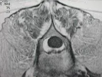
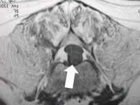
- Below left is an MRI of a spine with no herniated disc – note the black oval region is the spinal cord and the white halo surrounding the spinal cord is fat; below right is an MRI of a spine with a large amount of herniated disc material – note the flattened black oval region is the spinal cord and the black circular region immediately below the spinal cord is the herniated disc material which is compressing the spinal cord.