

Key Points
A free flap is a piece of tissue, usually skin or muscle that is transplanted from one area of the body to a wound that needs to be repaired
The blood supply of the tissue flap is reconnected to small blood vessels adjacent to the wound so that the flap stays alive
This type of surgery is an excellent treatment option for complex wounds in dogs
Indications for free tissue transfer
- Microvascular free tissue transfer is used for wounds that are caused by
- traumatic accidents
- oncological surgery (tumor removal) – see example below
- radiation therapy burns
- thermal burns
- chemical burns
- vesicant burns – injected medications such as chemotherapeutic medications or some injectable anesthetics
- The procedure has the advantage of requiring only one surgery to achieve a cosmetic result.
Types of free flaps that are used to repair wounds
- Skin flaps are used for wounds that have a relatively good blood supply
- Muscle flaps are used for wounds that do not have a good blood supply with exposed bone. Muscle has a phenomenal capability to remove infection and heal the wound. A skin graft is placed over the muscle.
Surgical procedure
- A normal segment of skin along with its nutrient artery and vein are removed from the donor site (inner thigh or side of shoulder). The blood vessels of the skin flap are surgically reconnected (with the aide of an operating microscope to visualize the vessels) to the blood vessels adjacent to the wound thereby allowing the skin flap to survive in its new location.

Case example – Medial Saphenous Fasciocutaneous Microvascular Free Flap
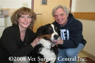
- Esther presented with a mast cell tumor located over the front of the wrist (called the carpus). Treatment options were amputation of the limb or excision of the tumor followed by radiation therapy. Because the tumor was fairly sizeable, a large section of skin needed to be removed in the process. A microvascular free flap is ideal in such cases, as this provides a durable covering over the wound that can withstand radiation therapy.
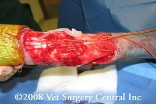
- In this video, a free flap is harvested from the inner thigh. The blood vessels that feed this portion of skin are also harvested with the flap and will be disconnected from the limb. A dog will function just fine without these saphenous vessels.
- In this video the artery of the skin flap is being sutured to an artery adjacent to the recipient wound. The artery pumps blood into the skin flap.
- In this video the vein of the skin flap is reconnected to a vein adjacent to the recipient wound. The vein drains blood out of the skin flap.

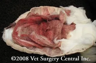
- Esther’s flap turned a purple color in the postop period. The photo right was taken on the third day after surgery. An instrument (doppler) used to test the blood flow in the flap indicated that the artery was still pumping blood in the flap. The problem was that blood was not flowing out of the flap very well due to swelling of the tissue which was compressing the veins. This uncommon complication can result in death of the flap unless appropriate treatment is provided.
- We therefore elected to use leech therapy to relieve the venous congestion. This entails placing the leeches directly on the flap (using a syringe casing) or pricking the skin with a needle and directly applying the leeches to the flap.
- After the leeches release themselves from the flap, the bite sites will continue to bleed which relieves congestion of the flap. Don’t be alarmed if the bandage is soaked with blood when it is changed (see photo right). Your doctor will monitor your pet for anemia if multiple leech therapy sessions are needed.
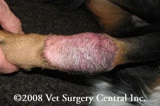
- Esther’s flap survived completely and the leeches helped it along. In the photo right, 13 days after surgery, the flap is healing well with the congestion and swelling ameliorated.
Success rate
- About 90 to 95 % of free tissue flaps survive transplantation.
Potential complications
- There is an inherent risk of anesthetic death with any procedure requiring anesthesia, however, this is very small.
- A blood clot could develop in the artery or vein of the flap at the site where they have been reconnected, which may lead to death of the entire flap.
- Venous congestion may occur due to swelling of the tissues which partially occludes the vein of the flap. In most cases this is reversible once the swelling starts to come down, however, in some cases some sutures may need to be removed to relieve compression of the tissues and blood vessels. Leech therapy may also be recommended in some cases.
- During the healing process the surgery site may become itchy and the dog may lick or chew at the site. If this occurs the vessels could become damaged and the flap may die.
- The site where the flap was collected from may break open if the dog is too active or is allowed to lick and chew at the region.
- A pooling of fluid under the area where the skin flap was removed can occur in a small percentage of patients undergoing this procedure, however the fluid will become absorbed over time.
- It is possible that the hair of the skin graft might grow in a different color or direction than the native hair coat around the region to be reconstructed.
Postop care
- Starting the evening prior to surgery, aspirin should be given to help reduce the risk of blood clot formation in the vessels of the flap. The dose of aspirin is 2 mg/kg twice daily for four days (equates to about one 81 mg tablet for an 80 pound dog).
- Bandage changes initially will be done daily to check the health of the flap; it is common that bandages are changed by the owners; bandages must not be put on tight as this could compress the blood vessels that feed the flap.
- It is imperative that the patient is prevented from self-mutilating the flap with the aid of bandages and/or Elizabethan collars for up to 6 weeks.
- Follow up checkups with your veterinarian are necessary to evaluate the progress of the skin flap.
References
- Miller JM, Lanz OI, Degner DA. Rectus abdominis free muscle flap for reconstruction in nine dogs. Vet Surgery 36:259-265, 2007.
- Degner, DA, Walshaw, R., Fowler, J.D. et al: Surgical Approaches to Recipient Vessels of the Fore and Hindlimbs for Microvascular Free Tissue Transfer in Dogs. Vet. Surg. 34 (4), 297–309, 2005.
- Degner, DA, Walshaw, R., Fowler, J.D. et al: Surgical Approaches to Recipient Vessels of the Head and Neck for Microvascular Free Tissue Transfer in Dogs. Vet. Surg. 33:200-208, 2004.
- Jackson AH, Degner DA, Myiawaki T, Jackson IT, Silverberg: The deep circumflex iliac free flap in cats. Vet Surg. 32 (4), 341–349, 2003.
- Myiawaki T, Degner DA, Eleszar H, Barakat K, Jackson IT: Easy tissue expansion of prelaminated mucosal-lined flaps for cheek reconstruction in a canine model. Plast Reconst Surg 109:1978-85, 2002.
- Calfee EF, Lanz OI, Degner DA, Peterson SL, Duncan RB, Broadstone RV, Martin RA, Austin B: Microvascular free tissue transfer of the rectus abdominis muscle in dogs. Vet Surg 31:32-43, 2002.
- Lanz OI, Broadstone RV, Martin RA, Degner DA: Effects of epidural anesthesia on microcirculatory blood flow in free medial saphenous fasciocutaneous flaps in dogs. Vet Surg 30:374-9, 2001.
- Bebchuk TN, Degner DA, Walshaw R, Brourman JD, Arnoczky SP: Evaluation of a free vascularized medial tibial bone graft in dogs. Vet Surg 29: 128-44, 2000.
- Robertson H, Degner DA, Walshaw R: Replantation of a partially amputated paw in a dog. Vet Comp Ortho Trauma 12:40-42, 1999.
- Fowler JD, Degner DA, Walshaw R: Microvascular free tissue transfer: Results in 57 consecutive cases. Vet Surg 27:406-412, 1998.
- Degner DA, Walshaw R: Medial saphenous fasciocutaneous and myocutaneous free flap transfer in eight dogs. Vet Surg 26:20-25, 1996.
- Degner DA, Lanz OI, Walshaw R: Myoperitoneal microvascular free flaps in the dogs: an anatomical study and clinical case report. Vet Surg 25:463-70, 1996.
- Degner DA, Walshaw R, Lanz OI, et al: The medial saphenous fasciocutaneous free flap in the dog. Vet Surg 25:105-113, 1996.
- Degner DA, Walshaw R, Arnoczky SP, Smith RJ, Degner LA, Hamaide A: Evaluation of the cranial rectus abdominis muscle pedicle flap as a blood supply for the caudal superficial epigastric skin flap. Vet Surg 25:292-299, 1996.
- Degner DA, Walshaw R, Kerstetter KK: Vascular anomaly of the prescapular branch of the superficial cervical artery and vein of an omocervical free skin flap in a dog. Vet Comp Ortho Trauma 8:102-106, 1995.
- Degner DA, Bauer MS, Steyn PF, Toombs W, Orton CE: The cranial rectus abdominis muscle pedicle flap in the dog. Vet Comp Ortho Trauma 7:21-24, 1994.
- Degner DA, Bauer MS, Steyn PF, Toombs W, Orton CE: The cranial epigastric skin flap in the dog. Vet Comp Ortho Trauma 7:18-20, 1994.
- Degner DA, Bauer MS, Cozens M: Reverse saphenous conduit flap: a case report in a cat. Vet Comp Ortho Trauma 6:175-177, 1993.
