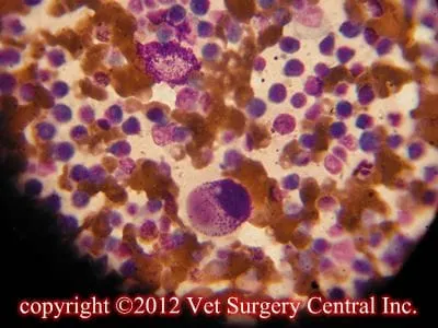
Key Points
Mast cell tumors are a lot larger than they appear to the naked eye, therefore large margins of normal looking tissue must also be removed
Mast cell tumors in cats and ferrets frequently are benign
Radiation therapy is an effective means to treat mast cell tumors that cannot be surgically removed
Chemotherapy is indicated to shrink tumors down to resectable size and for patients that have high grade tumors
What are mast cells?
Mast cells are normally found in the tissues of the body. They release histamine when stimulated by mechanical factors (tissue injury) or chemical factors (allergies). Histamine causes signs of allergies such hives and increases production of acid in the stomach. Proteolytic enzymes released by mast cells delay healing of incisions. Heparin, a blood thinning agent also found in the mast cells, increases the bleeding into traumatized tissues. There are many other chemical messangers that are found in mast cell granules, but these will not be discussed in this article.
Mast cell tumors
A mast cell tumor is a mass of cancerous mast cells. These tumors are common location for mast cell tumors is in the skin. Mast cell tumors can be located in the fatty layer beneath the skin. They can form in organs such as the liver, spleen, intestines, stomach. Theses tumors may spread into lymph nodes, the blood stream, the bone marrow or the internal organs.
Effects of mast cell tumor degranulation
Excessive levels of histamine in the blood stream from the mast cell tumor degranulation can cause stomach ulcers to develop. Signs of stomach ulcers include vomiting blood or coffee ground-like material, black stools, decreased or loss of appetite, and stretching in a praying position (due to abdominal pain). Extensive bruising and swelling is common following fine needle aspiration of high grade tumors. Shock and death may occur due to acute degranulation of the tumor cells. Poor healing of the incision following tumor excision, especially if residual tumor is left behind.
Signs of a mast cell tumor
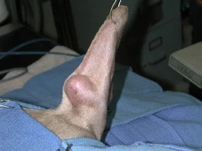
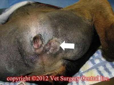
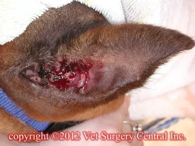
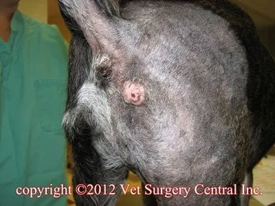
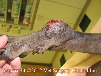
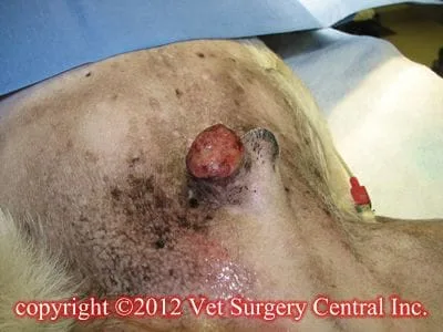
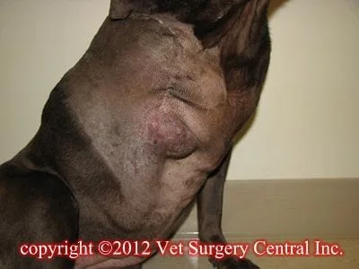
Mast cell tumors have been refered to as the “master of disguise”, as they can mimick the appearance of benign tumors. The most common presentation of a mast cell tumor is a lump in the skin (so called “button tumor”); about 10% of the patients will have or will develop multiple mast cell tumors in the skin. The mass may appear as a raised pink lump (see photo below). Mast cell tumors located in the fatty tissues (subcutaneous tissues) frequently feel like an ill-defined mass; higher grade tumors tend to have a more ill-defined margins. If the lump is massaged or rubbed it may swell or form hives….called the Darier sign; this is caused by the release of histamine from the mast cells.
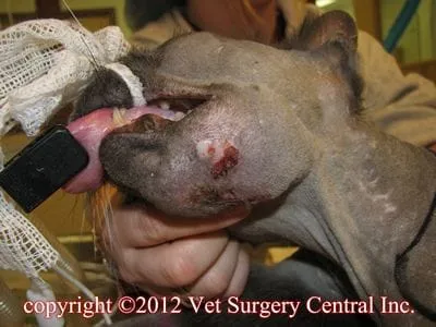
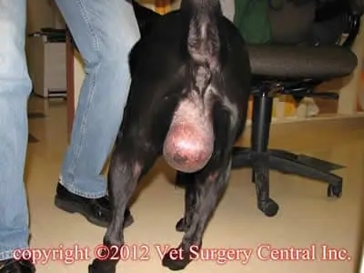
The photos below are from patients that had mast cell tumors.
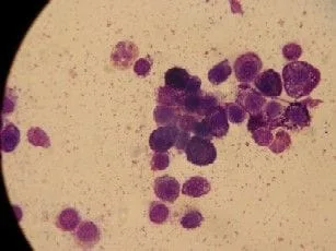
Diagnostic work-up
Surgery of mast cell tumors of the skin
Benadryl should be given to the patient prior to surgery to minimize degranulation effects of the mast cell tumors during surgery; high grade tumors are more prone to degranulating and causing low blood pressure or shock during surgery than mid and low grade tumors. In dogs, 2 cm of normal looking skin surrounding the tumor and a deep layer of tissue (dense fascia or muscle) should also be removed with the mass. High grade tumors should have 3 to 4 cm side margins and a deep layer of tissue removed around the mass. The removed tissue is evaluated by a pathologist to determine tumor type, tumor grade, and completeness of tumor removal.
In cats, mast cell tumors located in the skin are commoly grade 1 tumors, therefore only a small border (0.5 to 1 cm) of normal looking skin surrounding the tumor may be needed to be removed with the mass to achieve a surgical cure.
Additional therapy
If the histopathology report indicates that the mast cell tumor has incomplete or very close surgical margins, additional surgery can be performed to remove more tissue. This should be the first choice of treatment if additional tissue can be safely removed. The tissue again is sent to a pathologist to ensure that the entire tumor has been removed.
Chemotherapy may be recommended before or after surgery. Indications for adjunctive chemotherapy includes high grade tumors, high index of cell proliferation (proliferative study), cKit mutation, and presence of metastasis to regional lymph nodes, internal organs or the bone marrow. Prednisone, which is a steroid may help to shrink the tumor size, but is effective at best in only 20% of the cases. Incomplete response is due to prednisone does not kill the tumor cells rather decreases the size of the tumor by reducing tumor-associated inflammation; a positive response is typically seen for only 1 to 2 months. Vinblastine, vincristine, lomustine (CCNU), cyclophosphamide, hydroxyurea (chemotherapy) can improve the long-term survival in some patients. Chronic treatment with benadryl has been suggested for dogs that have developed multiple mast cell tumors. Palladia is a tyrosine kinase receptor blocker; the proliferative panel is used to determine if the patient should be prescribed this medication. The tumor should be positive for cKIT mutations for this drug to be potentially effective. Ulcers of the stomach and intestine have been a common side effect of this medication, but this may be due to overdosing this medication.
Radiation therapy may be used to “clean up mast cells that are left behind” following incomplete removal of a mast cell tumor. Mast cell tumors are moderately sensitive to radiation therapy. Radiation therapy is not a good choice if the tumor has spread to the internal organs.
Prognosis in dogs
In dogs, the prognosis following treatment of mast cell tumors of the skin is directly related to the grade of the tumor as determined by the biopsy results (histopathology). If the tumor has been completely removed with surgery, then recurrence of the tumor should be low. Grade 1 tumors have a benign behavior and an excellent cure rate with surgery. Grade 2 tumors bear a moderately malignant behavior, and have a 20% recurrence rate following aggressive surgery. Grade 3 tumors are very malignant, commonly metastasize and 10% of these patients are alive at 1 year following surgery.
Some pathologists subclassify grade 2 mast cells as high grade 2 or low grade 2 mast cell tumors; high grade 2 tumors behave like grade 3 tumors and low grade 2 mast cell tumors behave like grade 1 tumors. Beware that grading mast cell tumors is very subjective. In one study, mast cell tumors were graded by a group of pathologists and they commonly disagreed upon the grade of the tumors. A much better testing method of determining the how malignant or benign a mast cell tumor will behave is a proliferative study. This includes PCNA (proliferating cell nuclear antigen), AgNOR (agyrophilc nuclear oganizing regions), and Ki67. Tyrosine kinase receptors (a receptor for mast cell growth factor) is also important and tests related to this include cKIT mutations and KIT staining patterns. All of these tests in the panel are important, however cKIT mutations are very dangerous and the Ki67 also seems to be very important. These tests are more expensive than standard testing (histopathology), but they seem to be worthwhile.
Mast cell tumors located on the muzzle and oral cavity tend to have a more malignant behavior. Mast cell tumors located in the perineal region (anus, scrotum and vulva) may not have as aggressive of behavior as previously believed, however tumor in these locations may be more difficult to remove. Dogs that have evidence of mast cells also in the internal organs, blood stream or bone marrow have a poor prognosis.
Tumors that are ulcerated may result in a poor prognosis. Dogs that have signs of gastrointestinal ulcers (vomiting, black stools) generally have a poor prognosis. Tumors that are large have a poor prognosis (larger than 4 cm). Tumors that have shown recent rapid growth have a poor prognosis. Mast cell tumors which have not changed for a period of 2 months frequently have a good prognosis.
Tumors that have recurred after surgical removal may result in a poor prognosis. Dogs living greater than 30 weeks after surgery and not having any recurrence of local or distant tumor are considered cured
Prognosis in cats
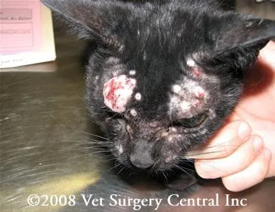
In cats, the prognosis following removal of mast cell tumors of the skin is usually excellent and surgery is commonly curative. The prognosis of mast cell tumors localized to the spleen is good and many live for an additional one to two years after the spleen is removed. Sometimes cats have multiple mast cell tumors all over their body which may spontaneously resolve or may progress with time and result in death (see photo below).