

Key Points
There are two main types of lung tumors: ones that start in the lung and ones that have spread from another part of the body to the lung
If there is a single lung tumor that originated in the lung, surgery is the best option for a potential cure
In selected cases of metastatic lung tumors, surgery will also be recommended
Types of lung tumors
- Metastatic lung tumors are those that spread from a primary tumor else where in the body
- example is a dog that has a bone tumor which has spread to the lungs
- metastatic lung tumors generally are usually multiple
- if your dog has multiple lung masses these also could be caused by a fungal infection and not cancer
- Primary lung tumors originate in the lung tissue
- the most common primary lung tumor is bronchoalveolar carcinoma
- the most common place that these tumors can spread to is the lung
Clinical signs of lung tumors
- Cough which may also produce phlegm or blood
- Loss of appetite
- Weight loss
- In the early stages, there may be no clinical signs, but your veterinarian may detect this tumor on chest radiographs
Testing done for patients in preparation for surgery
- Complete Blood cell count
- Chemistry profile
- Urinalysis
- Chest radiographs – left side, right side and front-back views
- tumors located in the right lung are seen best when the pet is lying on the left side and visa versa
- If fungal infections are suspected, fungal titers may be indicated
- In selected cases in which infection is suspected a scope (camera) may be placed down the wind pipe and into the lungs in order for samples to be collected for microscopic analysis and/or bacterial culture
- Fine needle biopsy of the lung mass is occasionally recommended
Typical findings on radiographs
- Primary lung tumors frequently are located in the caudal (towards the hind end of the pet) lung lobes, however can be located in any lung lobe; they are usually a single mass in the lungs, unless the tumor has spread
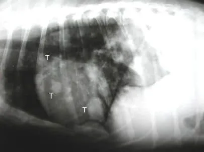
- Metastatic lung tumors frequently are multiple and found in a variety of lung lobes; occasionally a metastatic tumor spread from else where in the body may appear as a single mass in the lungs; if your pet has a fever and has been traveling to southern United States, the masses in the lungs could be due to fungal infection and may be treated with medication
- In the radiograph below left, take note of the multiple round masses in the lungs representing metastatic disease (only three of the many tumors were labeled T in this photo)
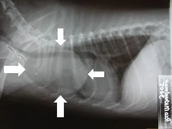
- In the radiograph below right, take note of the single mass located in one of the lung lobes; this is typical of a primary lung tumor
Metastatic lung tumors
- A diagnosis of cancer can only be definitively be made with a lung biopsy and histopathology
- If there is no evidence of a primary tumor in the body and multiple nodules are found in the lungs, a minimally invasive lung biopsy can be done by placing a thoroscope (camera) into the chest and obtaining a sample of lung; animals that have this done can usually go home the same day after the procedure; this test will confirm a diagnosis of fungal or cancer
- If there is a known tumor somewhere on the body and the lungs are full of nodules, it is presumed that these are metastatic tumors
- If there are three or fewer lung nodules, surgery can be done to remove these in order to increase the lifespan of your pet; this is most commonly done with osteosarcoma metastatic disease
Primary lung tumors
- Surgery is recommended for these cases if there is no evidence of metastatic disease
- If needed, the entire left lung can be removed at one time, as the right lung will take over for the left
- Your pet may not do as well if the entire right lung (all lobes) need to be removed
- Usually the tumor is removed through the side of the chest cavity (lateral thoracotomy)
- In some cases the breast bone must be divided (median sternotomy) so that both left and right lungs can be accessed
Aftercare following thoracotomy and lung tumor removal
- IN HOSPITAL
- Your pet is initially given oxygen via a nasal catheter
- Pain medication is given to your pet either by injection or via an epidural catheter, or through the intravenous as a constant rate infusion
- Antibiotics are usually just given at the time of surgery and not given in the postop period
- Intravenous fluids are given to keep your pet hydrated
- A bandage may be placed on the chest
- AT HOME
- Limit exercise to short leash walks for 3 weeks if a lateral thoracotomy was performed; limit exercise for 6 weeks if a median sternotomy was performed
- Administer oral pain medications as needed
- Monitor respirations to make sure that these are not labored
- Monitor gums and tongue to make sure that these are pink
- Encourage eating
- Monitor the incision for signs of infection
- Do not allow licking or scratching of the incision
- Put a tee shirt on your pet to help prevent this
- Chemotherapy may be recommended by an oncologist, pending the tumor type
Potential complications
- Anesthetic death – uncommon
- Seroma formation at the incision – fluid accumulation which will resolve within about 3 to 4 weeks
- Infection – uncommon
- Internal hemorrhage – uncommon
- Spread of tumor – dependant on type and stage of tumor
- Pneumothorax – air leaking from lung after surgery – uncommon, but requires that a chest tube be kept in place for a longer period of time
