

Key Points
- Single large liver tumors frequently are the kind that have a low tendency to spread to other parts of the body and are amenable to surgery
- Liver tumors can cause nonspecific signs such as vomiting, weight loss, decreased appetite, lethargy and pale gums
- Patients having surgery frequently have a good prognosis, providing that the tumor is self-contained and the surgeon can completely remove the tumor
Types of liver tumors
Metastatic liver tumors are those that spread from a primary tumor else where in the body example is a dog that has a tumor of the spleen which has spread to the liver. Metastatic liver tumors generally are usually manifested as multiple masses within the liver. With that said, multiple liver masses they could just be benign nodular change (hyperplastic regenerative nodules) which has a good prognosis and does not require surgical removal.
Primary liver tumors originate from the liver tissue and the most common primary liver tumor is the hepatocellular carcinoma. These tumors have a low tendency to spread, but invade into the liver tissue.
Clinical signs of liver tumors
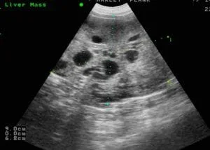
Vomiting, loss of appetite, weight loss, yellow color of the skin and eyes (jaundice) and lethargy are potential clinical signs of the liver tumors. Most patients that present to us have no obvious clinical signs of illness, and the liver mass may be incidentally discovered on x-rays or ultrasound being performed for as a routine screening diagnostic or found when a pet is investigated for elevation of liver enzymes.
Testing done for patients in preparation for surgery
Blood work in the form of a Complete Blood cell count, Chemistry profile and Coagulation profile are baseline tests that are commonly completed. Abdominal ultrasound is commonly done to initially diagnose the present of a liver mass; however, CT scan of the chest and abdomen is our advanced imaging modality to define the borders of the tumor, determine which vessels are feeding the mass and to rule out spread of the tumor to other parts of the body. Fine needle biopsy of the liver mass is occasionally recommended if there is concern for a type of cancer called lymphoma which is a type of tumor that is treated with chemotherapy.
Metastatic liver tumors
- A diagnosis of cancer can only be definitively be made with a liver biopsy and histopathology
- If there is no evidence of a primary tumor in the body and multiple nodules are found in the liver, a minimally invasive liver biopsy can be done by placing a laparoscope (camera) into the abdomen to obtain a sample of liver; animals that have this done can usually go home the same day after the procedure; this test will confirm a diagnosis of cancer
Primary liver tumors
- Surgery is recommended for these cases if there is no evidence of metastatic disease
- If needed, more than half of the liver can be safely removed and the liver will regenerate
- Usually the tumor is removed through an incision made on the midline of the abdomen
- In some cases the breast bone must be divided (median sternotomy) so that the tumor can be accessed
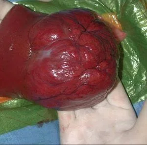
- Even tumors that seem to invade deep into the liver can be safely removed using a technique that we developed called the hilar liver dissection technique (see reference below). With this technique, liver tumors can be removed with much less bleeding and with a better chance that all of the tumor has been completely removed. The procedure does not require fancy equipment such as the “Cavitron Surgical Aspirator”, just a sound knowledge of the vascular anatomy of the liver. The video (right) demonstrates the resection of a tumor located on the left side of the liver.
Aftercare following liver tumor removal
- IN HOSPITAL
- Pain medication is given to your pet either by injection or via an epidural catheter, or through the intravenous as a constant rate infusion
- Antibiotics are usually just given at the time of surgery and not given in the postop period
- Intravenous fluids are given to keep your pet hydrated
- AT HOME
- Limit exercise to short leash walks for 3 weeks if a midline abdominal incision was performed; limit exercise for 6 weeks if a median sternotomy was performed
- Administer oral pain medications as needed
- Monitor respirations to make sure that these are not labored
- Monitor gums and tongue to make sure that these are pink
- Encourage eating
- Monitor the incision for signs of infection
- Do not allow licking or scratching of the incision
- Put a tee shirt on your pet to help prevent this
- Chemotherapy may be recommended by an oncologist, pending the tumor type; hepatocellular carcinomas usually are not too responsive to chemotherapy and surgery is usually the only treatment needed
Potential complications
- Anesthetic death – uncommon
- Seroma formation at the incision – fluid accumulation which will resolve within about 3 to 4 weeks
- Infection – uncommon
- Internal hemorrhage – uncommon
- Spread of tumor – dependant on type and stage of tumor
References
- Covey J, Degner DA, Jackson AH, Hofeling A, Walshaw R. Hilar liver resection in dogs. Accepted for publication. Vet Surgery 2008
- Liptak, JM: Hepatobiliary tumors, in Withrow SJ, Vail DM (eds): Small Animal Clinical Oncology (ed 4). St. Louis, WB Saunders, 2007, pp 483-485
- Bachellier P, Ayav A, Pai M, et al: Laparoscopic liver resection assisted with radiofrequency. Am J Surg 193:427-430, 2007
- Tobias KM: Surgical stapling devices in veterinary medicine: A review. Vet Surg 36:341-349, 2007
- Gayet B, Cavaliere D, Vibert E, et al: Totally laparoscopic right hepatectomy. Am J Surg 194:685-689, 2007
- Saiura A, Yamamoto J, Koga R, et al: Usefulness of LigaSure for liver resection: Analysis by randomized clinical trial. Am J Surg 192:41-45, 2006
- Liptak JM, Dernell WS, and Withrow SJ. Liver tumors in cats and dogs. Comp Cont Ed Vet 26:50-57, 2004
- Martin RA, Lanz OI, Tobias KM: Liver and Biliary System, in Slatter DH (ed): Textbook of Small Animal Surgery (ed 3). Philadelphia, PA, Saunders 2003, pp 716-717
- Morgan, P: A novel technique for parenchymal division during hepatectomy. Am J Surg 181:236-237, 2001
- Nuzzo G, Giulante F, Giovannini I, et al: Liver resection with or without pedicle clamping. Am J Surg 181:238-246, 2001
- Takayama T, Makuuchi M, Kubota K, et al: Randomized comparison of ultrasonic vs. clamp transection of the liver. Arch Surg 136:922-928, 2001
- Jarnagin WR, Gonen M, Fong Y, et al: Improvement in perioperative outcome after hepatic resection: analysis of 1, 803 consecutive cases over the past decade. Ann Surg 230:309-321, 1999
- Fan ST, Lai EC, Lo CM, et al: Hepatectomy with an ultrasonic dissector for hepatocellular carcinoma. Br J Surg 83:117-120, 1996
- Trout NJ, et al: Surgical Outcome of hepatobiliary cystadenomas in cats: five cases (1988-1993). JAVMA 206: 505, 1995
- Gozzetti G, Mazziotti A, Grazi GL, et al: Liver resection without blood transfusions. Br J Surg 82:1105-1110, 1995
- Lewis DD, Bellenger CR, Lewis DT, et al: Hepatic lobectomy in the dog—a comparison of stapling and ligation techniques. Vet Surg 10:221-225, 1990
- Bjorling DE, et al: Partial hepatectomy in dogs. Compend Contin Educ Pract Vet 3:257, 1985
- Francavilla A, et al: Liver regeneration in dogs: Morphologic and chemical changes. J Surg Res 25:409, 1978.
- Sleight DR, Thomford NR: Gross anatomy of the blood supply and biliary drainage of the canine liver. Anat Rec 166: 153-160, 1970
