

Key Points
A disease of the hip joint that results in abnormal deformity of the ball of the hip joint
This is a disease primarily seen in small breed dogs
Treatment is femoral head and neck excision
Prognosis is very good, providing that rehabilitation therapy is done after surgery
Same disease but different names (synonyms)
- Calve-Perthes disease
- Legg-Perthes disease
- Avascular necrosis of the femoral head
- Coxa plana
- Osteochondritis juvenilis
What is it?
- A disease of the hip joint that results in abnormal deformity of the ball of the hip joint
- The disease starts with an insult to the blood supply to the head of the femur bone
- As the blood supply is damaged, the bone dies off
- The bone in the femoral head collapses and the cartilage coating of the femoral head becomes cracked and deformed
- Arthritis or inflammation of the hip joint results, which causes pain and lameness
Cause
- Not completely understood
- Genetics – can be autosomal recessive gene in some dogs
- Injury – compression of the vessels of in the femoral head
- Abnormal sex hormone activity – preconcious
- Because of the potential heritability of this disease, affected dogs should not be bred
Signalment
- Typically dogs that are less than 1 year of age (5 to 8 months of age is most common with peak incidence around 7 months of age)
- Breeds commonly affected
- Yorkshire Terriers – most common breed
- Westies
- Many other small breed dogs
- Equal risk for males versus females
Clinical signs
- Irritability
- Chewing at hip of flank region
- Progressive lameness (may take about 2 months until dog is not bearing any weight on limb)
- Stiffness of affected limb (85 to 90% of dogs have only one hip affected)
- Atrophy of muscles of affected limb
- Pain when moving the hip – especially when extending the limb backward and also to the side
- Crepitus or crunchy feel of the hip joint on range of motion
Diagnosis
- Physical examination – hip pain
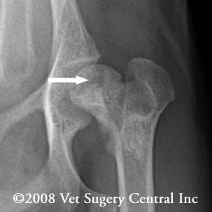
- Radiographs
- flattening of femoral head
- lucencies of femoral head
- bone spurs – arthritic degeneration
- increased joint space
- Below is a radiograph of the hips of a Yorkie that has the disease; take note of the moth-eaten appearance of the femoral head (below left and right)

Treatment
- Conservative treatment with strict cage rest and physical therapy could be successful only if the femoral head is still has its normal shape and is is tightly seated in the socket; monthly radiographs should be made until the pet is 1 year of age
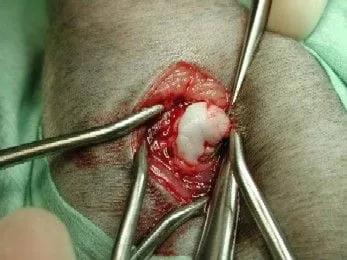
- Surgery – femoral head and neck excision is the treatment of choice if the radiographs show deformity of the femoral head or looseness of the hip joint; this surgery involves removing the femoral head and neck
- Below is a photo as the femoral head is being removed from the hip joint shown above; take note of the deformed head of the femur and the cracks in the cartilage. The radiograph below demonstrates a femoral head and neck excision (arrow)

Prognosis
- Femoral head and neck excision usually gives a satisfactory result – the pet is greatly improved; some dogs may have residual lameness, especially with heavy exercise or weather changes
Postop care
