

Key Points
Dogs should be 16 weeks of age to benefit from the surgery
Your pet can be screened to at 4 months of age using PennHip x-rays to determine if he/she is likely going to develop hip dysplasia
The JPS surgery has minimal morbidity and is usually done on an outpatient basis
There is about a 85% chance that affected dogs will benefit from having the procedure done
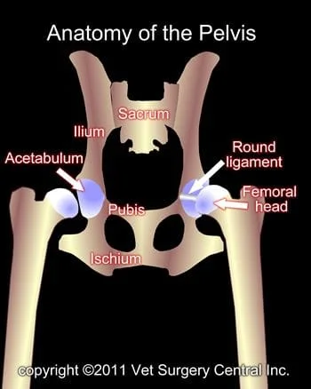
The pelvis is made of four bones: the ilium, acetabulum, pubis, and ischium. In the immature animal, these bones are not fused together, but as the pet matures, these bone fuse together in one confluent bone. The ilium joins the lower part of the spine called the sacrum.
The hip joint consists of a ball (femoral head) which is at the end of the femur bone and socket (acetabulum). This joint joins the hind limb to the pelvis. The joint is held together with a very strong round ligament (sometimes called the teres ligament) and the joint capsule. The muscles that surround the hip joint also provide very good support to the joint.
W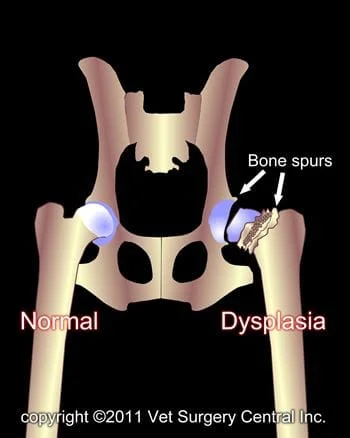
HIP = coxofemoral joint
DYSPLASIA = abnormal development
Hip dysplasia is an inheritable orthopedic condition, meaning that defective genes are passed from the parents to their offspring. In affected puppies, the soft tissues that support the hip are looser than normal and the muscles that support the hip are poorly developed. Some suggest that there is disproportionate development of the bones versus the muscles (i.e. bones grow too fast and muscle development is retarded). As a result, the femoral head slips in and out of the joint (called subluxation) when the puppy is ambulating. Because the bones of puppies are very soft, the subluxation of the joint causes the acetabulum, which is normally a deep cup to become shallow like a saucer. The femoral head, which is a round ball, assumes flattened shape like a mushroom. In addition, the “roof” of the acetabulum can develop painful microscopic fractures from the subluxating femoral head. Secondary arthritis develops in the hip joint which results in lameness, stiffness, and pain.
Signs
Dogs show signs of hip dysplasia from 5 to 8 months of age, but some dogs are in their early adult years or during the geriatric stage of life. Commonly affected breeds include German Shepherds, Labrador retrievers, Golden retrievers, Rottweilers and many other large breed dogs. Small breeds also develop this problem with Pugs and cocker spaniels commonly affected. The main clinical signs include lameness, bunny hopping when running, difficulty getting up after rest, reluctance to exercise, and sometimes crying due to severe pain.
Identification of candidates for the JPS surgery
Dogs should be 16 weeks of age for this surgery to be beneficial. Most dogs at this age do not have any clinical signs of hip dysplasia. As a result, breeds such as Labradors, Rottweilers, German Shepherds, Golden retrievers and other susceptible breeds should be tested at 16 weeks of age using PennHIP radiographs should be made to identify dogs that have loose hips.
Preparation for surgery
The pet should be fasted prior to surgery, as instructed by the surgical team. Water is usually permitted up to the time of admission to the hospital. An antacid such as Pepcid AC may be prescribed and should be administered by 6 AM on the day of surgery; this treatment will help reduce the risk of esophagitis (heartburn) in the postop period. The surgical team should be informed of any medications that your pet is currently receiving. The pet should not receive any aspirin within 1 week of surgery, as this medication will thin the blood and increase the risk of bleeding. Just prior to surgery, your pet will receive a sedative, have an intravenous catheter placed for the administration of intravenous fluids and intravenous medications, be induced under general anesthesia with medication(s), and have a breathing tube (endotracheal tube) placed to allow delivery of oxygen and gaseous anesthesia. The surgical site will be clipped and cleansed with an anti-septic solution in preparation for surgery. While under general anesthesia, the pet’s breathing will be assisted with a ventilator and vital parameters such as heart rate, respiratory rate, core body temperature, blood pressure, oxygenation of the blood (pulse oximetry), exhaled carbon dioxide (capnography), and heart rhythm (EKG) will be monitored to ensure the pet’s well being. Pain will be controlled both during and after surgery with analgesics (pain-controlling medication). Please note that each surgical and anesthesia team may elect to chose a different, but effective analgesia protocol.
JPS surgery
Your pet will be put under general anesthesia. A small incision is made between the hind limbs to expose the pubic bone of the pelvis. The growth plate of the pubis is cauterized to destroy the growing cells of this part of the pelvis. Your pet likely will go home on the same day that the surgery is performed
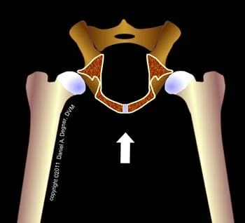
The illustration of the pelvis (right) is viewed from a dog’s rear end and the back part of the pelvis has been removed for demonstration purposes. The growth plate (growth center) of the pubic bone (arrow) is destroyed with electrocautery. Thus, this portion of the pelvis no longer grows. The remaining parts of the pelvis grow, which rotates the acetabulae (sockets of the hips) over the femoral head (ball of the hip). Place your cursor over the image to see the effect of the JPS surgery and continued pelvic growth. The result is stable hips that have much less chance of developing significant arthritis.
Success of the JPS procedure
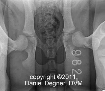
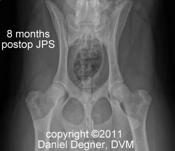
If the procedure is done at 16 weeks of age (or younger), there is a much greater chance that it will prevent significant development of arthritis of the hips, versus having the procedure done when the dog is older. Dogs that have a distractive index that is greater than 0.7 will have progressive arthritis and the JPS may not be a good procedure for such patients; generally, dogs that have a DI that is 0.4 to 0.7 will have a good chance to be helped with the JPS surgery. See photo below left shows loose hips on the distraction x-ray of a Standard poodle at 16 weeks of age and the photo below right is an x-ray showing very tight hips as a result of the JPS pulling the hips into the sockets. This patient is pain free and likely will not develop any arthritis of the hips.
Potential complications
Infection following this surgery is an uncommon complication. The most common complication is seroma or fluid accumulation at the surgery site and typically presents as a soft swelling at the surgery site; this fluid build up will resolve on its own in a couple of weeks. The urethra may be damaged (cauterized) during the procedure and results in urinary difficulties; this complication is rare. Progressive debilitating arthritis is a potential complication, but this problem is less likely to occur if the dog is a good candidate for the procedure.
rev 10/7/11
