Instability of the Hock Joint in Dogs and Cats

Key Points

Hock joint instability is a very debilitating condition that is caused by trauma

Instability can be due to tearing of ligaments or a fracture of the fibula or bottom of the tibia bone

Surgical repair of the ligaments or bones is needed for the best outcome

The prognosis tends to be better if the bone is broken and can be repaired versus a ligament tear alone


Anatomy

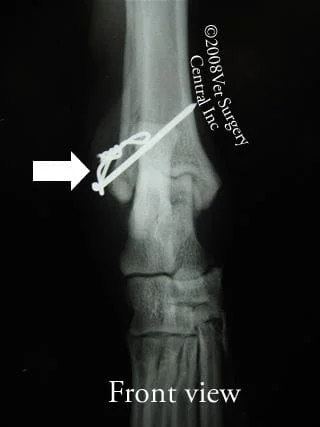
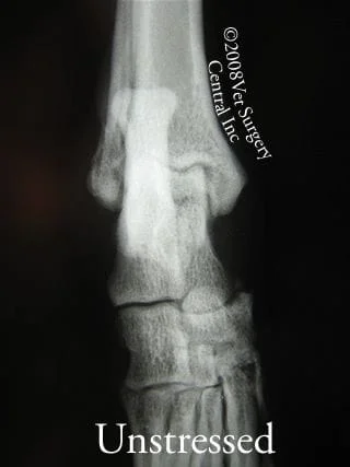
The hock joint of a dog or cat is equivalent to our ankle joint. The shin (tibia) bone is connected to the hock joint which then joins the talus bone (one of the bones of the paw). The fibula bone (splint bone) runs along the full length of the tibia bone. Ligaments on the inner and outer part of the hock joint hold the bones together. Each side of the hock joint has two important ligaments (total of four main ligaments that hold the hock together)

Signs

Owner will note acute lameness of the affected hindlimb and may note swelling of the hock joint; heat may be detected from the inflamed joint in dogs that do not have a dense hair coat. Hock instability is easily detected on physical examination. The veterinarian must test the hock by trying to open the joint from the outside and inside (medial and lateral) sides of the joint which the hock in extension and flexion. In addition, the hock must be stressed by twisting it to the inside and outside (internal and external torsion) to see if the ligaments are stable. The video to your right demonstrates hock instability; take note of the instability when the hock is twisted.

Diagnosis

preop hock rads It is important to determine the cause of instability of the hock joint prior to surgery. The hock will be put through a series of stress x-rays; that means that forces will be applied to the joint in a variety of directions and x-rays (radiographs) of the hock will be made. Take note of the normal appearing hock joint of a dog that has the instability as seen in the video above. Place your cursor over the image to discover the problem. The arrow denotes the fracture in the fibula bone which resulted in instability. The take home lesson from this case was that without stress radiographs the diagnosis would have been missed and the inappropriate treatment provided.


Treatment

postop_fib_fx_repair Treatment of hock instability is dictated by the type of injury that is present: ligament damage versus a fracture. If a facture is present surgery is recommended. Take for example the case discussed above has a fracture of the fibula bone. The treatment in this case was stabilization of the fracture using a pin and figure-of-eight cerclage wire. Alternatively, a screw could be placed. In order to minimize the development of arthritis in the joint, the fracture must be pefectly aligned and the pin must not penetrate the joint. If a ligament is torn, screws are placed in the bones above and below the joint and heavy permanent suture is tied around screws to simulate the ligament. With time scar tissue develops over the hock joint to add support to the surgically repaired joint.


Contact Us

We look forward to hearing from you

Office Hours

24 Hour Patient Care MON 8:00AM - FRI 6:00PM

Flint Office

Monday

8:00 am - 6:00 pm

Tuesday

8:00 am - 6:00 pm

Wednesday

8:00 am - 6:00 pm

Thursday

8:00 am - 6:00 pm

Friday

8:00 am - 6:00 pm

Saturday

Closed

Sunday

Closed

Contact Us

Flint Office

Monday
8:00 am - 6:00 pm
Tuesday
8:00 am - 6:00 pm
Wednesday
8:00 am - 6:00 pm
Thursday
8:00 am - 6:00 pm
Friday
8:00 am - 6:00 pm
Saturday
Closed
Sunday
Closed
Contact Us

Location

Find us on the map