

Key Points
Hip dysplasia is the abnormal development of the hip joints and usually has a genetic basis.
A variety of medications are available to temporarily relieve pain associated with the hip joints. Surgery, however, frequently is needed.
Femoral head and neck excision is usually reserved for small patients; triple pelvic osteotomy is used for immature dogs that have minimal to no arthritic changes of the hips; total hip replacement is a salvage procedure that is used in large breed dogs.

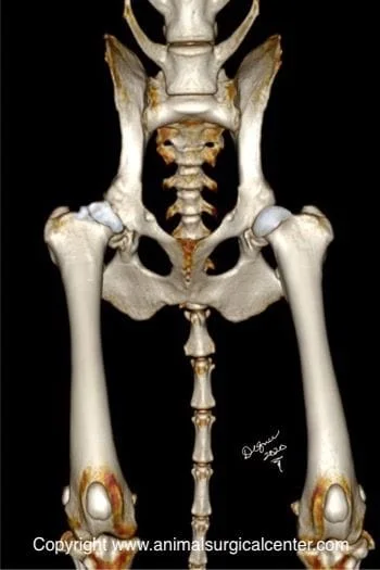
The pelvis is made of four bones: the ilium, acetabulum, pubis, and ischium. In the immature animal, these bones are not fused together, but as the pet matures, these bone fuse together in one confluent bone. The ilium joins the lower part of the spine called the sacrum.
The hip joint consists of a ball (femoral head) which is at the end of the femur bone and socket (acetabulum). This joint joins the hind limb to the pelvis. The joint is held together with a very strong round ligament (sometimes called the teres ligament) and the joint capsule. The muscles that surround the hip joint also provide very good support to the joint.
HIP = coxofemoral joint
DYSPLASIA = abnormal development
Hip dysplasia is an inheritable orthopedic condition, meaning that defective genes are passed from the parents to their offspring. In affected puppies, the soft tissues that support the hip are looser than normal and the muscles that support the hip are poorly developed. Some suggest that there is disproportionate development of the bones versus the muscles (i.e. bones grow too fast and muscle development is retarded). As a result, the femoral head slips in and out of the joint (called subluxation) when the puppy is ambulating. Because the bones of puppies are very soft, the subluxation of the joint causes the acetabulum, which is normally a deep cup to become shallow like a saucer. The femoral head, which is a round ball, assumes flattened shape like a mushroom. In addition, the “roof” of the acetabulum can develop painful microscopic fractures from the subluxating femoral head. Secondary arthritis develops in the hip joint which results in lameness, stiffness, and pain.
Signs
Dogs show signs of hip dysplasia from 5 to 8 months of age, but some dogs are in their early adult years or during the geriatric stage of life. Commonly affected breeds include German Shepherds, Labrador retrievers, Golden retrievers, Rottweilers and many other large breed dogs. Small breeds also develop this problem with Pugs and cocker spaniels commonly affected. The main clinical signs include lameness, bunny hopping when running, difficulty getting up after rest, reluctance to exercise, and sometimes crying due to severe pain.
Diagnosis
A diagnosis of hip dysplasia is based on a combination of history, clinical signs, physical examination findings, and radiographs. Below is a set of radiographs from a dog that had hip dysplasia. The radiograph below left was made when the dog was immature, versus the radiograph below right was made a few years later (no treatment provided). Take note of the loose hips and the severe arthritis that has developed.


Preparation for surgery
The pet should be fasted prior to surgery, as instructed by the surgical team. Water is usually permitted up to the time of admission to the hospital. An antacid such as Pepcid AC may be prescribed and should be administered by 6 AM on the day of surgery; this treatment will help reduce the risk of esophagitis (heartburn) in the postop period. The surgical team should be informed of any medications that your pet is currently receiving. The pet should not receive any aspirin within 1 week of surgery, as this medication will thin the blood and increase the risk of bleeding. Just prior to surgery, your pet will receive a sedative, have an intravenous catheter placed for the administration of intravenous fluids and intravenous medications, be induced under general anesthesia with medication(s), and have a breathing tube (endotracheal tube) placed to allow delivery of oxygen and gaseous anesthesia. The surgical site will be clipped and cleansed with an anti-septic solution in preparation for surgery. While under general anesthesia, the pet’s breathing will be assisted with a ventilator and vital parameters such as heart rate, respiratory rate, core body temperature, blood pressure, oxygenation of the blood (pulse oximetry), exhaled carbon dioxide (capnography), and heart rhythm (EKG) will be monitored to ensure the pet’s well being. Pain will be controlled both during and after surgery with analgesics (pain-controlling medication); we routinely place an epidural catheter for administration of narcotics during surgery and for two days after surgery. Please note that each surgical and anesthesia team may elect to chose a different, but effective analgesia protocol.
Treatments
A variety of medications are available to temporarily treat pain associated with the hip joints. Surgery, however, frequently is needed. There are three surgical procedures that may be recommended. If your pet does not have arthritis in the joint a reconstructive procedure (triple pelvic osteotomy) can be done to make the socket fit better over the ball of the femur bone. If your pet already has arthritis one of two procedures is recommended: femoral head and neck excision or total hip replacement. The femoral head and neck excision involves cutting off the ball and neck of the femur so that it does not rub against the socket of the hip. In this situation, the body forms a false joint and the pain is relieved. This procedure is recommended for medium and small dogs and cats. Large and giant breed dogs may not do as well with this type of surgery. Total hip replacement is recommended for the large and giant breeds of dogs. This procedure involves replacement of the hip socket with a polyethylene cup and replacing the head of the femur with a metal implant.
Medical treatments
A number of nonsurgical measures can be taken to help alleviate signs of hip dysplasia and these include achieving an ideal body weight (weight loss for most pets), low impact regular exercises (swimming is excellent), provision of a a warm, soft bed (infrared beds may be helpful), avoidance of cold flooring such as cement floors, administration of pain relieving medication (tramadol, nonsteroidal anti-inflammatories), administration of high levels of omega fatty acids (Joint diets or Welactin), administration of glucosamine/chondroitin products, extra-corpreal shock wave therapy, therapeutic laser, acupuncture, and rehabilitation therapy by a professionally trained therapist.
Success rate of the surgery
Total hip replacement is about 90% successful. In large breed dogs femoral head and neck excision usually does not return the function of the limb to perfectly normal, but can significantly decrease the pain that the pet is experiencing. Cats and small dogs do very well with this procedure. Triple pelvic osteotomy is about 90% successful, however, some arthritis may develop, as the pet gets older.
Potential complications
HIP REPLACEMENT. Infection at the surgical site can occur soon after surgery or months to years later. If your pet gets an infection following total hip replacement, usually the implants will need to be removed. To prevent infection from developing, your pet should be administered antibiotics prior to and following any surgical procedure or dental work. This will help prevent bacteria from entering the blood stream and getting into the cement of the prosthesis. Following hip replacement, the prosthetic hip can become dislocated if your pet is too active during the healing phase. Too much activity can cause the cup or the femoral prosthesis to loosen from the bone, thus requiring a second surgery.
FEMORAL HEAD AND NECK EXCISION. Infection is an uncommon, but potential complication. Second, if your pet does not get enough exercise after surgery the hip will lose mobility and the false joint does not form properly. This will result in poor limb function after healing has taken place. Therefore rehabilitation therapy is very important.
PELVIC OSTEOTOMY. Infection is an uncommon, but potential complication. If your pet is too active during the first 2 months after surgery, the implants securing the pelvis can loosen and the repair may break down. Some hip arthritis will develop in most dogs receiving a triple pelvic osteotomy surgery, but usually does not cause pain or stiffness. If the hips develop debilitating arthritis total hip replacement may be needed.
rev 9/25/11
