

Key Points
Stomach bloat is a serious and potentially fatal disease, if early treatment is not instituted
Emergency decompression of the stomach and surgical tacking of the stomach to the body wall is necessary
Survival rate following surgery is about 90% of the stomach is not devitalized. If a portion of the stomach has died off, the survival rate is about 50%, in spite of medical and surgical treatment
A laparoscopic preventative surgery should be done in breeds that are susceptible to this condition
Introduction
- The stomach is located in the front part of the abdomen and is the first part of the gastrointestinal tract located in the abdominal cavity. Gastric volvulus and dilatation (GDV) is a condition that starts off with distention of the stomach with food, fluid such as water, or air due to excessive swallowing. The stomach twists in a clockwise direction as it becomes distended. The inlet to the stomach from the esophagus and the outlet leading into the duodenum get kinked off and the food, fluids and air cannot pass in either direction. As a result, unproductive retching occurs.
- Due to the twisting and displacement of the stomach, the blood supply may be cut off and a portion or all of the stomach may die. The longer that the stomach has been twisted before treatment is provided, the greater the risk that the stomach will die.
- Another event that occurs is occlusion of the main vein (vena cava) leading from the back half of the body to the heart and resultant shock. Shock, a condition in which there is inadequate perfusion of the body with blood, is fatal if not treated. Signs of shock include pale gums, rapid heart rate, and weak pulses (low blood pressure). Dogs that develop GDV generally are quite ill and need immediate aggressive treatment.
- Infrequently the nerves to the stomach get damaged from the GDV and the paralysis of the muscles of the stomach wall occurs. This results in chronic bloating that is usually not responsive to any therapy.
- Left is an illustration of a stomach in normal position (E=esophagus; D=duodenum); right – after the stomach twists 270o in a clockwise direction, the duodenum as it exits the stomach, is moved to the top and behind the stomach, the esophagus is twisted and the stomach is becomes distended like a balloon.
Treatment of GDV
- Initially intravenous catheters are placed and fluids are administered to help reverse shock. Next a sedative is given and a tube is passed into the stomach. The stomach is pumped if needed to remove any air, fluid, or undigested food. If the tube cannot be passed into the stomach due to the twisting of the esophagus, a large needle is passed through the side of the abdomen into the distended stomach.
- The pet is then taken to surgery. The internal organs are examined for damage. The stomach is repositioned and it is tacked to the right side of the abdominal cavity to prevent a future GDV. Sometimes a portion of the stomach is found to be dead and needs to be removed. If too much of the stomach is dead euthanasia may be recommended. If the stomach is filled with food or other material that cannot be removed with a tube, which is passed down the throat, the stomach is opened up and the ingesta is removed.
- Occasionally the spleen is also twisted and the blood clots have developed in its vessels. In this case the spleen will be removed (splenectomy). Dogs can live normal lives without a spleen. There is about a 40% chance that the heart will develop an abnormal rhythm if a dog has also developed a problem with the spleen that mandates splenectomy. The abnormal heartbeat may require treatment to prevent death.
- While the pet is in the intensive care unit, careful monitoring is done in order to ensure that the internal organs are functioning properly. Fluid therapy, medications, and transfusions (plasma and/or packed red cells) may be needed after surgery.
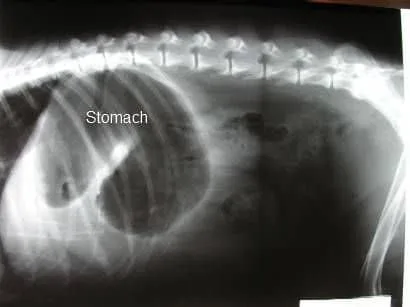
- Figure 1 below – side view of a dog’s abdomen: take note of the stomach is moderately distended with gas in a dog with GDV. The stomach has a reverse “C” shape which indicates that it is twisted
- Figure 2 below: the belt loop gastropexy involves creating a belt or tongue of tissue on the stomach.
- Figure 3 below: a belt loop is created in the muscle wall of the abdomen on the right side.
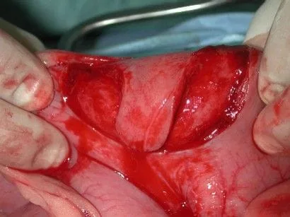
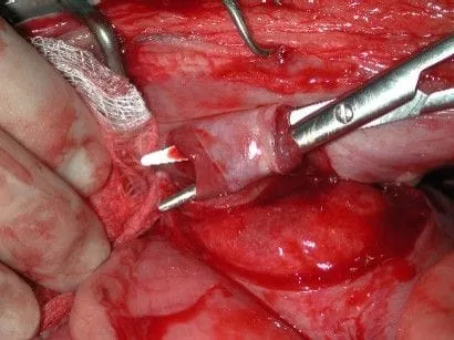
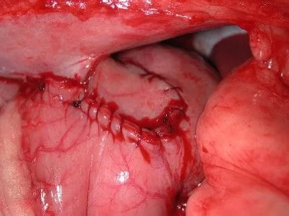
Figure 4: the belt or tongue of tissue created on the stomach has been pulled through the beltloop of the abdominal wall and the belt is sutured onto the stomach, completing a beltloop gastropexy.
After care and convalescence
- After surgery has been completed intensive care must be provided for the best chance for a successful outcome. Intravenous fluids and antibiotics are administered for at least 24 hours. If the blood protein level is low, artificial plasma (Hetastarch) is administered. Blood pressure is carefully monitored and maintained.
- Electrocardiograms are monitored after surgery, as abnormal heartbeats called arrhythmias are common. Usually the arrhythmias will show up within the first 24 hours. Sometimes these need to be treated with medication, as they can be fatal, however, is not common.
- Your pet will be monitored for disseminated intravascular coagulation (DIC). In this situation the clotting system becomes hyperactive and small blood clots develop in the internal organs and multiple organ failure occurs. Generally DIC is fatal when it occurs, therefore early treatment is the only potential safeguard.
- In general, about 90% of the dogs having GDV, if treated early will survive. By 10 to 14 days after surgery most patients are felling very well. If a portion of the stomach is dead and needs to be removed, the survival rate is about 50%.
- Restricted activity should be instituted so that the tacking of the stomach does not break down. The pet should be kept on a leash when taken outside, and no running, jumping, or horseplay is permitted. By 3 to 4 weeks out for surgery the healing is complete for the most part.
- After a pet has had GDV, the daily ration of food should be divided into 2 to 3 meals and exercise should be restricted for 2 hours after eating to prevent rebloating (the stomach will not twist because of the gastropexy); rebloating is not common problem in dogs following surgery.
Potential complications
- As with any surgery, complications may arise. Even though relatively uncommon, anesthetic death can occur. With the use of modern anesthetic protocols and extensive monitoring devices (blood pressure, EKG, pulse oxymetry, inspiratory and expiratory carbon dioxide levels, and respiration rate), the risk of problems with anesthesia is minimal.
- Infection is also an unusual complication as strict sterile technique is used during the surgery and antibiotics are administered in the perioperative period.
- Abnormal heartbeats called arrhythmias
- Disseminated intravascular coagulation results in dysfunction of multiple internal organs, bleeding disorder, and commonly death.
- Breakdown of the stomach tack (gastropexy) occurs less than 5% of the time. Recurrent GDV can occur.
- Chronic recurrent bloat. Recurrent bloat generally occurs infrequently, but usually is due to very poor function of the muscle of the stomach. Medication may be administered to improve the stomach motility, but this will work in only about 50% of the patients. The presentation of this is very similar to a GDV, but the stomach does not twist. A gastropexy (stomach tack) can also breakdown and result in recurrent bloat and twisting of the stomach, but this is unusual (less than 5% chance). Surgery is done to correct this problem.
- Because of the high variability of the status of patients that have GDV, it is also very difficult to predict whether your pet is going to have postoperative complications. In general, dogs having necrosis of a portion of the stomach do not fair as well as those having minimal trauma to the stomach.
Prognosis
- If treated early, about 90% of the dogs having surgery will survive
- If at the time of surgery, a portion of the stomach was found to be dead, the dog has a 50% chance to survive
- A preventative surgery can be performed to minimize the risk of GDV in high risk patients such as Great Danes, German Shepherds, Blood Hounds, Irish Setters, Irish Wolfhounds, Standard Poodles, and other susceptible breeds. Great Danes are at very high risk and research has shown that one in four Danes will bloat in their lifetime. This surgery is done laparoscopically via two small incisions with the aid of a camera. The surgery can be done as early as 6 months of age (at the time of neutering). This procedure has minimal morbidity, less anesthesia and surgery time, short hospital stay (done as an outpatient surgery), and is less expensive than treating GDV.
