

Key Points
A Gartner cyst is an embryonic male reproductive structure in female animals that should not have formed
Removal of this structure is generally easy, but critical knowledge of the anatomy is essential for a complication free outcome
Prognosis is excellent
Anatomy
The reproductive tract in the fetus has both male and female parts. The female part is called the Mullerian ducts system and the male part is called the Wolffian duct system. Due to hormonal influences, the male duct system disappears in females and visa versa in males. Sometime a portion of the Wolffian system remains in female animals (and humans). Because this duct is lined with mucus producing cells, it produces fluid. This duct system is located between the vagina and the cervix. The duct has no opening into the vagina and no opening into the uterus, therefore it becomes a cystic, closed structure. With time the cyst enlarges with fluid and can compress internal organs such as the bladder, urethra (tube that leads to the outside from the bladder) and colon. If this structure gets infected, the patient may become very ill
Signs
In some patients the Gartner cyst may not cause clinical signs until it has reached a critical size. In symptomatic patients, straining to urinate or defecate may be the most common signs. The veterinarian usually will be able to feel this cyst with a rectal examination and potentially with abdominal palpation. If the cyst becomes infected, the patient will become depressed, may have a fever, have abdominal pain and may vomit.
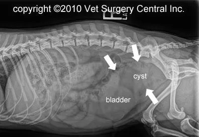
The diagnosis of this condition is based on abdominal x-rays which may show a mass like structure within the abdomen above the bladder. The photo right shows a Gartner cyst that is compressing the bladder. Ultrasound is also useful to identify identify the structure has a Gartner cyst. Ultimately, findings at surgery and biopsy of the structure provide a definitive diagnosis.
Preparation for surgery
Make sure that your pet is fasted, as instructed by your pet’s surgical team. Water is usually permitted up to the time of admission to the hospital. Your pet’s surgeon may prescribe an antacid such as Pepcid AC, which should be administered by 6 AM on the day of surgery; this treatment will help reduce the risk of esophagitis (heartburn) in the postop period. Inform the surgical team of any medications that your pet is currently receiving. Your pet should not receive any aspirin within 1 week of surgery, as this medication will thin the blood and increase the risk of bleeding. Prior to surgery, your pet will receive a sedative, have an intravenous catheter placed for the administration of intravenous fluids and intravenous medications, be induced under general anesthesia with medication(s), and have a breathing tube (endotracheal tube) placed to allow delivery of oxygen and gaseous anesthesia. The surgical site will be clipped and cleansed with an anti-septic solution in preparation for surgery. While under general anesthesia, your pet’s breathing will be assisted with a ventilator and vital parameters such as heart rate, respiratory rate, core body temperature, blood pressure, oxygenation of the blood (pulse oximetry), exhaled carbon dioxide (capnography), and heart rhythm (EKG) will be monitored to ensure your companion’s well being. Pain will be controlled both during and after surgery with analgesics (pain-controlling medication).
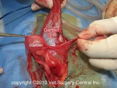
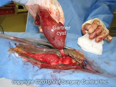
Removal of Gartner cyst is a relatively easy, but delicate procedure. The peritoneum (lining over the cyst) is opened up and the cyst is gently freed up. Care is taken to prevent damage the ureters (tubes that come from kidneys and enter the neck of the bladder), bladder, ureters, nerves that enter the neck of the bladder (for urinary continence) and the colon.
Video of Gartner cyst removal in a dog
