

Key Points
By definition, a gallbladder mucocele is an accumulation of thick mucus within the gallbladder
Surgical removal of the gallbladder is typically recommended
Short-term prognosis is fair, however patients surviving the perioperative period have excellent long-term survival rates
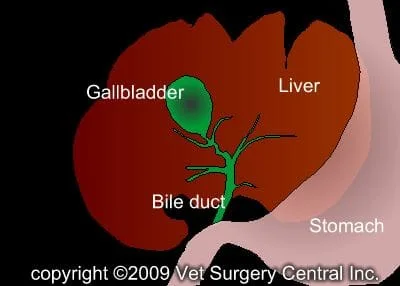
The liver contains very small bile channels (tubes) called canaliculi that collect bile from the liver cells. These ducts join larger and larger ducts that ultimately terminate in 3 to 5 visible ducts that empty bile into the gallbladder. The common bile duct (technically called the “bile duct”) is the final stretch of tubing that delivers bile into the first part of the small intestine. The gallbladder is nestled between two liver lobes on the right side of the liver. Bile, which is concentrated by the gallbladder, serves as a vehicle for fat absorption. Cholecystokinin, a hormone that is released from the pancreas when food is consumed, causes the gallbladder to contract and expel its contents into the intestine via the bile duct.
Cause of gallbladder mucoceles
Historically, gallbladder diseases were rarely diagnosed in dogs and cats. With the advent of advanced imaging tools such as ultrasound, the diagnosis of gallbladder disease is commonly recognized in dogs and cats. Unlike humans, dogs and cats rarely develop stones in the gallbladder. A mucocele is the most common condition that afflicts the gallbladder. By definition, a mucocele is an accumulation of thick mucus within the gallbladder. Infection is infrequently associated with a mucocele. This condition occurs as a result of excessive proliferation of the mucus-producing cells within the lining of the gallbladder. Thick mucus cannot be expelled from the gallbladder. As the gallbladder becomes distended, its blood supply is impaired and it subsequently is prone to rupturing. Once ruptured, bile will leak into the abdomen from the gallbladder and cause the patient to become severely ill.
Signs
Gallbladder mucocele is more prevalent in mid-aged to older medium-sized breeds of dogs such as Cocker Spaniels, Miniature Schnauzers and Shetland Sheepdogs. Most dogs with a mucocele have nonspecific signs such as vomiting, loss of appetite and lethargy. Other signs that may be found on a physical examination include abdominal pain, jaundice, elevated respiratory rate, fever and elevated heart rate.
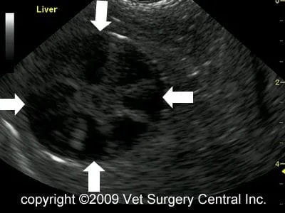
Bloodwork commonly will frequently show elevation of liver enzymes with this condition. With rupture of the gallbladder, liver enzymes are usually very elevated and the white blood cell counts are mild to markedly elevated. Ultrasound is the “gold standard” to diagnose the problem and commonly reveals a “kiwi” appearance of the gallbladder (see photo).
The day of surgery
Emergency surgery will be recommended if the gallbladder is ruptured. If the patient is stable and the gallbladder is still intact, surgery may be scheduled the next day. If your companion has elective gallbladder surgery, make sure that your companion has been fasted prior to surgery and that the prescribed dose of Pepcid AC has been administered on the morning of the procedure. The surgeon will contact you after the surgery to give you a progress report after the surgery. Our anesthesia and surgical team will prescribe a pain management program, both during and after surgery that will keep your companion comfortable. This will include a combination of general anesthesia, injectable analgesics, and oral analgesics.
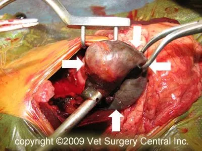
Medical therapy may be recommended in select cases; however, surgery is typically needed in most cases. Surgical removal of the gallbladder is the treatment of choice. In some cases, a stent (rubber tube) may be placed in the common bile duct, to ensure continued flow of bile. Following surgery, the patient will receive pain-relieving medication to ensure a comfortable recovery. Intravenous fluids will be administered to ensure that your companion remains hydrated after surgery. Blood testing will be performed after surgery to monitor your companion’s recovery.
Results
The perioperative mortality ranges from 22 to 32%; however, rupture of the gallbladder has been shown to worsen the prognosis when infection is present. Long-term survival of patients that have undergone gallbladder removal is excellent. Liver enzymes remain elevated in most patients, but these values usually are much lower after the healing process is completed.
Aftercare
At home, the incision should be checked for signs of infection. Your companion should not lick the incision, as this could open the incision or cause infection. If necessary, an Elizabethan collar can be placed on your companion to prevent licking and chewing at the surgical site. A low fat diet may be recommended in some patients; however, a regular diet may be tolerated. Antibiotics may be prescribed after the surgery if bacterial infection is suspected or confirmed by culture results. Exercise should be restricted for about 3 weeks after surgery to allow uncomplicated healing of the incision.
References
- Aguire AL, Center SA, Randolph JF, et al. Gallbladder disease in Shetland Sheepdogs: 38 cases (1995–2005). J Am Vet Med Assoc 231:79–88, 2007.
- Amellem PM, Seim HB, MacPhail CM, et al. Long-term survival and risk factors associated with biliary surgery in dogs: 34 cases (1994–2004). J Am Vet Med Assoc 229:1451–1457, 2006.
- Mehler SJ, Mayhew PD, Drobatz KJ and Holt DE. Variables associated with outcome in dogs undergoing extrahepatic biliary surgery: 60 Cases (1988–2002). Veterinary Surgery 33:644–649, 2004.
- Pike FS, Berg J, King NW, et al. Gallbladder mucocele in dogs: 30 cases (2000–2002). J Am Vet Med Assoc 224:1615–1622, 2004.
- Worley, DR, Hottinger Lawrence HJ. Surgical management of gallbladder mucoceles in dogs: 22 cases (1999–2003). J Am Vet Med Assoc 225:1418–1422, 2004.
- Neer TM. A Review of disorders of the gallbladder and extrahepatic biliary Tract in the dog and cat. Journal of Veterinary Internal Medicine 6:186-192,1992.
