

Key Points
Recurrent urinary tract infection in female dogs may be due to excessive folds of skin that cover a female dog’s privates
A nip-and-tuck type of surgery may be very beneficial in controlling urinary tract infections
Healing time is about 2 weeks after surgery
Anatomy
The vulvar lips are folds of skin located on your female dog’s back side and is the enterance to the vestibule (cavity or room) that connects to the vagina. The opening of the urethra (where urine is expelled) is located at the front part of the vestibule, just behind the opening of the vagina. Skin folds are located over the top and sides of the vulva.
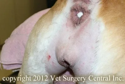
Excessive folds
Some female dogs have excessive skin folds that form a hood over the vulva. Early spaying of a dog may result in a small vulva that is tucked beneath surrounding skin folds. Obesity also will result in excessive tissue that tucks the vulva inward. The result is a persistant moist environment in the skin folds, which promotes excessive growth of bacteria in this area. The bacteria, which are mobile (can swim) move into the vestibule, up the urethra and into the bladder. In addition, the moisture can result in scalding of the skin of the vulva, thus cause your pet to have discomfort and irriation in this region.
Clinical signs
The primary clinical sign of excessive vulvar folds may be recurrent episodes of urinary tract infection. Signs of urinary tract infection may include frequent urination, urgency to urinate, urinary accidents in the house, straining to urinate, and a brown or bloody discoloration of the urine. Rubbing the hindend on the carpet or floor, licking the vulva, and crying in pain may be signs of ulceration of the skin of the vulva (urine scald and dermatitis). Excessive skin folds covering the vulva is sometimes very obvious, and other times needs the judgement of a veterinarian.
Your veterinarian will determine if your dog has excessive folds of skin that are covering the vulva. A urinalysis and urine culture along with blood testing (complete blood count and chemistry profile) is completed to evaluate the funtion of the kidneys and other internal organs and to check for urinary tract infection. Additional diagnostic testing includes x-rays and ultrasound of the abdomen. Endoscopy (camera is inserted to see the inside of the urinary tract, vestibule and vagina) are performed to check for anatomical abnormalities that could be causing urinary tract infection. In some cases, a dye study of the urinary tract with x-ray or CT scan may be recommended.
The Day of Surgery
In preparation for surgery, your pet should be fasted starting at 10 PM the night before surgery, however water does not need to be with held. To help prevent heartburn after surgery, a single dose of Pepcid AC (10 mg tablet per 20 pounds of body weight) should be administered at 6 AM at home on the day of surgery. Our anesthesia and surgical team will prescribe a pain management program, both during and after surgery, that will keep your companion comfortable. This will include a combination of general anesthesia, epidural catheter (for repeated injections of morphine), and oral analgesics.
Treatment
Episoplasty involves removing the folds of skin from around the vulva. This patient has the lines depicting the skin incisions drawn on the skin, which resembles horse-shoe shape. This patient is photographed as seen above, but the skin is pulled back to expose the vulva. In addition, the surgeon will remove excessive fat from this area.
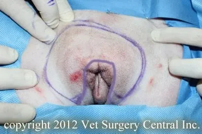
The skin is sutured to complete the procedure.

Outcome
Many patients are helped with this procedure. In general, this surgery is an under utilized procedure for the control of chronic recurrent urinary tract infections. Other underlying causes also may predispose to urinary tract infections, thus this procedure is not always effective in all cases.
