

Key Points
Ectopic ureter is an abnormally located terminal portion of the ureter. Instead of the ureter opening in the bladder, it opens in the urethra, vagina, or uterus. The result is constant dribbling of urine.
Surgical intervention is required to treat this condition
Prognosis is variable and but surgery could be curative
Medical therapy may also need to be provided after surgery if continued incontinence is present
Anatomy
- The kidney is made of a many very small filters called nephrons
- The final filtrate from the nephrons ultimately becomes urine
- The nephrons drain into a collecting chamber in the kidney called the renal pelvis
- The tube that carries urine from the kidney to the bladder is called the ureter
- The tube that carries urine from the bladder to the outside is called the urethra
Definition
- Ectopic ureter is an abnormally located terminal portion of the ureter. Instead of the ureter opening in the bladder, it opens in the urethra, vagina, or uterus. The result is constant dribbling of urine.
Signs of an ectopic ureter
- Urinary incontinence (leakage of urine)
- Blood tinged urine if bladder infections are also present
- Frequent urination
- More common in large breed dogs such as Labrador Retrievers; very rare condition in cats
- Almost exclusively diagnosed in females
Urinary incontinence (leakage of urine) and ectopic ureters
- In a dog that has an ectopic ureter, urine flows down the ureter directly into the urethra instead of the bladder (see illustration below). As a result, these pets typically drip urine constantly. Because this condition is congenital, most of these dogs are incontinent when they are born. The problem may be first noticed after the puppy has been weaned from its mother, because she may constantly clean the puppy.
- Urinary continence is also maintained by a valve at the neck of the bladder called the urinary sphincter. About 50% of the dogs that have an ectopic ureter have an abnormally weak urinary sphincter, and are also incontinent due to this (urinary sphincter mechanism incontinence).
Diagnostic testing prior to surgery
- Complete blood cell count
- Biochemical profile to make sure that the kidneys and other internal organs are healthy
- Urinalysis and urine culture to check for infection of the bladder and or kidney
- Abdominal ultrasound to evaluate the structure of the kidneys; the ureters usually cannot be seen with ultrasound unless they are dilated
- An excretory urogram involves giving the pet a type of dye or contrast media through an intravenous catheter. The dye is excreted into the urine. As the dye passes through the ureters they may be seen with radiographs (x-rays)
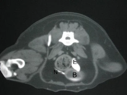
- CT scan is the best diagnostic tool used to diagnose ectopic ureters. This scan is performed if the excretory urogram does not provide a definitive diagnosis. Below is a CT scan image (this cross-section is through the lower part of the bladder at the level of the green arrow in the illustration above) of a normal ureter (labeled N) entering the bladder (labeled B) and a dilated ectopic ureter (labeled E).
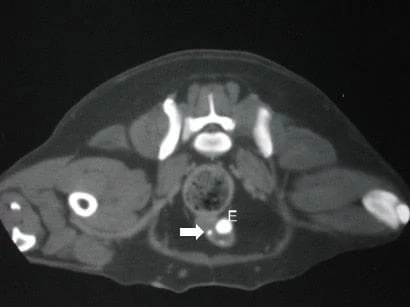
- The CT scan below shows a dilated ectopic ureter (labeled E) located within the urethral wall (this cross-section is at the level of the red arrow in the illustration above); also take note of the the smaller ectopic ureter labeled with an arrow
Surgery
- An incision is made along the midline of the abdomen
- The urinary system is examined and the abnormal ureter identified
- An incision is made in the bladder and first part of the urethra in order to perform the intricate work
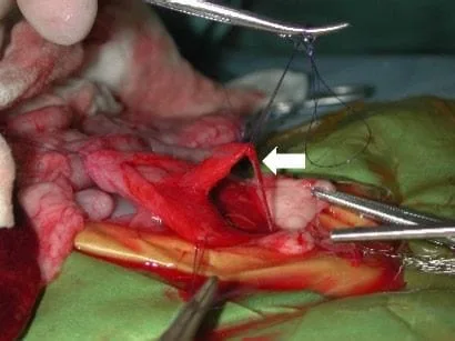
- The ectopic ureter is dissected out of the wall of the urethra (see below) and sutured into the bladder
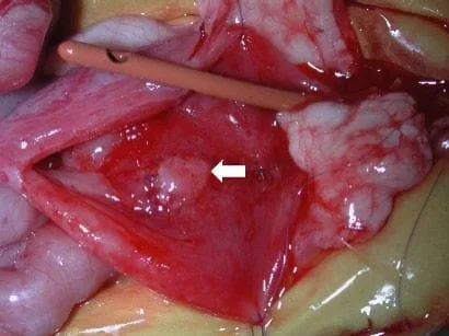
- The region where the ureter was dissected out of the urethra is sutured, which tightens the valve of the bladder. In the photo below the new opening of the ureter is sutured in the bladder
- A colposuspension surgery is performed in order to improve the tone of the valve of the bladder; this involves creating a sling around the urethra by suturing the front part of the vagina to the body wall, around the urethra.
- In the event the kidney belonging to the ectopic ureter is nonfunctional, it will be removed. This should not affect the lifespan of your pet, providing that the other kidney is normal
Complications
- Anesthetic death
- Allergic reaction to the dye
- Bladder infection
- Abdominal infection
- Continued incontinence
- Straining to urinate due to irritation of the bladder (resolves within the first 2 weeks)
- Obstruction of the urethra due to swelling, which may require temporary catheterization
Prognosis
- In our experience, about 75% of the dogs will have normal urinary continence after this surgery; the literature reports a lower success rate, however, these reports do not include cases that received the triad of surgeries discussed above: ureteral replantation, urethroplasty, and colposuspension
- In some cases medication (phenylpropanolamine) may be required after surgery to achieve complete urinary continence
