

Key Points
The most common type of tumors of the toe is squamous cell carcinoma and melanoma
Surgical amputation of the affected toe is the treatment of choice
Prognosis generally is very favorable for squamous cell carcinoma and fair for melanoma
Anatomy
The digit is commonly known as the toe. Each digit has 3 phalanges bones. The nail bed is attached to the third phalange bone.
Digital tumors
Tumors of the digits (toes) most commonly include squamous cell carcinoma, melanoma, osteosarcoma, hemangiopericytoma, benign soft tissue tumors and malignant soft tissue tumors. Squamous cell carcinoma accounts for more than 50% of all digital tumors. This tumor, which originates from skin cells, is very locally invasive and commonly will destroy the bone in the digit (see photo below left). Melanoma, the second most common tumor, accounts for 16% of all digital tumors. These tumors originate from the pigment-producing cells in the skin called melanocytes. These cells are responsible for giving humans a tan with sun exposure. Melanomas of the nail bed spread rapidly to other areas of the body. At the time of diagnosis of a digital melanoma, one-third of all affected dogs will have detectable spread to the lungs. Melanomas of the digits that do not involve the nail bed and are confirmed to be benign on the biopsy usually do not metastasize (see photo below right).


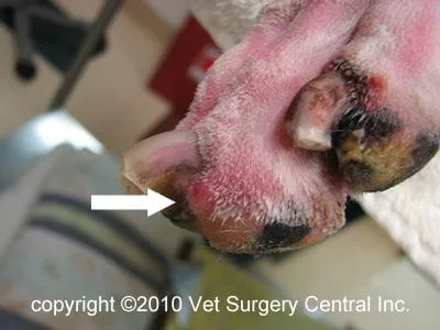
Squamous cell carcinoma is most commonly seen in large breed dogs with black coats. Over-represented breeds include Labrador Retrievers and Standard Poodles. Digital tumors will cause the toe to swell (see photo right) and may cause lameness. Initially, a tumor of the digit may mimic the appearance of an infected toe; however, treatment with antibiotics does not resolve the problem. If the tumor appears to be darkly pigmented, a melanoma is more probable; however, some melanomas lack pigment and may mimic the appearance of a squamous cell carcinoma. Enlargement of lymph nodes in the area of the tumor may be a sign of spread of the tumor to these nodes. If the tumor has spread to the lungs, potential clinical signs may include breathing difficulty, coughing, weight loss, poor appetite and malaise.
Diagnosis
The diagnosis of a digital tumor is based upon a fine needle biopsy or surgically collecting a piece of tissue from the mass. If the fine needle biopsy does not provide a definitive diagnosis, a core of tissue may be required. A complete blood count, chemistry profile and urine testing are done to evaluate the health status of your companion’s internal organs prior to anesthesia and surgery. X-rays of the affected digit may show destruction of the bone, especially if the tumor is a squamous cell carcinoma. Chest x-rays are used to help rule out spread of the cancer to the lungs and lymph nodes in the chest (see x-ray right which shows spread of melanoma tumors to the lungs). Abdominal ultrasound is also performed to rule out spread of tumor to the internal abdominal organs. Lymph nodes in the area of the tumor are aspirated to rule out spread of the cancer to the nodes, regardless of whether they are enlarged or not. If this test does not provide a clear-cut answer, removal of a regional lymph node and analysis of the node by a pathologist is recommended.

Our anesthesia and surgical team will prescribe a pain management program, both during and after surgery that will keep your companion comfortable. This will include a combination of general anesthesia, injectable analgesics, local anesthetics, oral analgesics and anti-inflammatory medication. The surgeon will call you with a progress report following the surgical procedure.
Treatments
Surgery is essential to treat a digital tumor. If the tumor is located on the toenail bed, the entire toe must be amputated. In some cases, benign tumors of the skin of the digits may be removed without removing the digit. Adjunctive therapy (chemotherapy or radiation) may be indicated for some malignant tumors. Usually surgery is all that is needed for a squamous cell carcinoma, however, melanomas should be treated more aggressively with a melanoma vaccination, chemotherapy and/or radiation.
An oncologist may recommend chemotherapy for your companion. Typically, one treatment is administered every 3 weeks for a total of 4 to 6 treatments. Most patients tolerate the chemotherapy medication with transient mild side effects.
Radiation therapy has been shown to prevent or delay the onset of tumor regrowth. Eighteen to 21 radiation treatments are administered to the tumor site and regional lymph nodes, starting 2 weeks after the tumor has been removed. Radiation treatments are administered Monday through Friday with no treatment during weekends. A short anesthesia is required during administration of each radiation treatment.
Aftercare
After surgery, a prescribed pain reliever should be given to minimize discomfort. Exercise must be limited for three weeks after surgery so that uncomplicated healing can take place. For one week, a bandage likely will be placed on the operated limb to protect the incision. Thereafter, the incision should be checked daily for signs of infection. Two weeks after surgery, the surgeon will monitor the healing process and our oncologist will initiate radiation therapy if indicated by the biopsy report. The oncologist may also recommend chemotherapy in selected cases. Melanoma vaccine can be administered as soon as a diagnosis of melanoma has been established.
Results
Digital melanomas: Digital melanomas treated with surgical amputation of the digit have a median survival time of 365 days. Digital melanomas that are not located on the nail bed and have benign characteristics (low mitotic index) on the biopsy are commonly cured with surgery alone. Inoculation with the melanoma vaccine can provide excellent long-term control of the disease and increased survival times for melanomas. Bergman reported only minimal to no side effects, which at worst was mild local reaction at the injection site. The best result is seen with intradermal vaccination that must be administered with a special injector system (see photo right of a intradermal vaccination). Squamous cell carcinoma: In a series of 21 cases, only 1 dog had local recurrence of the tumor and another dog developed metastasis of the tumor to the lungs after surgery. Therefore, this tumor has a very good chance to be cured with surgery alone.
Short-term complications following surgery are uncommon and may include temporary dehiscence (opening) of the incision and infection. Tumor recurrence and spread of cancer are other complications. Rarely, amputation of a digit will cause ongoing lameness on the operated limb.
References
- Henry CJ, et al. Canine digital tumors: a veterinary cooperative oncology group retrospective study of 64 dogs. J Vet Intern Med. 2005 Sept-Oct;19(5):720-4.
- Bergman PJ, et al. Development of xenogeneic DNA vaccine program for canine malignant melanoma at the Animal Medical Center. Vaccine. 2006 May 22;24(21):4582-5.
- Bergman PJ, et al. Long-term survival of dogs with advanced malignant melanoma after DNA vaccination with xenogenic human tyrosinase: a phase 1 trail. Clin Cancer Res 2003 Apr; 9(4):1284-90.
- Marino DJ, et al. Evaluation of dogs with digit masses: 117 cases (1981 -1991). J Am Vet Med Assoc. 1995 Sep 15;207(6):726-8.
- O’Brien MG, et al. Treatment by digital amputation of subungual squamous cell carcinoma in dogs: 21 cases (1987 – 1988). J Am Vet Med Assoc. 1992 Sep1;201(5):759-61.
