

Key Points
This surgical procedure is used to alleviate pain in a joint that has end-stage arthritis or other severe debilitating disease of a joint
The joint is permanently fused by removing the cartilage of the joint surfaces and placing bone graft in the joint; the joint is stabilized using metal implants so that the bone can heal together
Most dogs do well following surgery
Introduction
- Hyperextension (joint extends well beyond what is normal) can tear the ligaments that support the carpus (wrist) and result in a devastating instability of the joint that in almost all cases will not heal unless the injury is minor.
- The palmar fibrocartilage is the main supporting structure of the carpus in dogs and cats. If this structure is torn the pet will walk with the paw dropped down (hyperextended).
- The cause of hyperextension injury is typically a single traumatic event or repeated hyperextension of the carpus. Jumping off an elevated surface (out windows, off a roof, out of a pick-up truck, off a deck etc) or hitting the wrist against a solid surface (such as in fly ball) are the primary causes of this injury.
Diagnosis
The diagnosis of joint instability typically should include stress radiographs. This is performed by applying a stress to the joint and seeing where the damage has occurred. This is very important when evaluating damage to the carpus (wrist) or the tarsus (hind paw) in order for the veterinarian to determine which joints need to be fused. Place your cursor over the image; the two arrows show the separation of the back part of the joint indicating that the lower joint of the carpus is damaged, hence only a partial fusion of the joint is needed.
Treatment options
- A conservative approach usually is not successful in most cases as ligaments usually do not heal very well. In addition, if the behavior of the pet is not modified (i.e. the dog continues to jump off the deck onto the ground) reinjury is common. Conservative treatment includes supporting the affected limb in a splint for two months and restricting activity for three months.
- Surgery is generally the best option for a carpal hyperextension injury. The surgeon must first determine which which joint(s) of the carpus are unstable (see diagnostics above), then the surgeon can determine whether a partial or a complete fusion of the wrist joint is needed. Fusion of the unstable joints takes over the function of the torn fibrocartilage: to limit hyperextension of the wrist. More over, the fusion surgery will stop the pain that the dog has in the carpal joint.
- Partial fusion of the carpus results in normal to near normal function of the wrist. Complete fusion of the carpus will result in a wrist that has no movement at all. This still has a good outcome and many dogs actually can walk very well in spite of the loss of movement of the carpus.
Surgical principles
- All cartilage surfaces of the joint are removed
- Bone graft harvested from the patient is packed into the joints to stimulate healing
- Joint is positioned in a functional angle and stabilized with rigid fixation devices such as
- plates and screws
- screws
- pins and wires
- external skeletal fixator
- we prefer the use of plates and screws as this results in our best outcomes; we usually use an arthrodesis plate specifically made for this purpose
- The plates and screws usually are not removed.
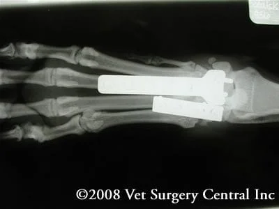
- Right is an example of a partial carpal arthrodesis in a dog. After healing had taken place the dog was able to walk once again without pain. Take note of the plate and screws that are holding and joints in a fixed position.
- Because a partial arthrodesis is performed the patient will have excellent function of the carpus with minimal function deficits.
Postop care
- Pain management
- The limb is supported in a cast or splint during the healing phase which is about 6 weeks
- The padding of the cast or splint is changed every 2 weeks
- Limited leash walks for urination/bowel movements until arthrodesis has been confirmed to be healed on radiographs
- Radiographs are taken 6 weeks after surgery
- After the cast has been removed, exercise is gradually increased on a leash over the next 6 weeks; during the first week a 5 minute walk twice daily is permitted; the walks can be increased by 5 minute increments each week until a normal amount of walking has been achieved
Potential complications
- Anesthetic death is very uncommon with our sophisticated monitoring devices and advanced anesthesia protocols
- Infection is possible but very uncommon
- Cold sensitivity requiring removal of the plate and screws after a year
- Failure of healing, necessitating regrafting of the joints
- Breakage of the plate or screws
- Pressure sores from the cast or splint
