

This condition occurs when the long digital extensor tendon tears out of the femur bone
Following surgery lameness typically resolves within 1 to 2 months
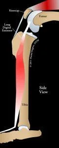
Tendons connect muscles to bones. The long digital extensor tendon attaches into the bottom of the thigh bone (femur), runs through the knee joint and attaches to its muscle located along the side of the shin bone (tibia).
Cause of long digital tendon avulsion
Generally this type of condition occurs in very young large breed dogs. At this age, the bones are very soft, thus the long digital extensor tendon can tear out of the bone with minimal trauma. Although not proven, it is quite possible that the patient may have a form of osteochondrosis (incomplete calcification of the bone in the region of the tendon), which is a weak spot at the attachment of the extensor tendon.
Signs
Breeds most commonly affected include Great Danes, Golden retrievers and Labrador retrievers. Dogs develop this problem when they are typically under 1 year of age; however, two reports have cited Great Danes that were 14 and 16 months of age when clinical signs of the problem were first noted. Clinical signs of this condition include lameness and swelling on the outer side of the knee, over the region of the avulsed tendon.
Diagnosis
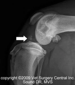
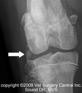
The diagnosis of a long digital extensor avulsion is supported by x-rays and physical examination findings. Uncommonly, CT scan or MRI of the knee may be needed to arrive at a diagnosis. Exploratory surgery is the most definitive way to diagnose the problem and treat the condition. In preparation for surgery, preoperative blood work including a complete blood count, chemistry profile and urine testing are recommended to ensure that your pet is healthy to under go anesthesia and surgery.
Preparation for surgery
The pet should be fasted prior to surgery, as instructed by the surgical team. Water is usually permitted up to the time of admission to the hospital. An antacid such as Pepcid AC may be prescribed and should be administered by 6 AM on the day of surgery; this treatment will help reduce the risk of esophagitis (heartburn) in the postop period. The surgical team should be informed of any medications that your pet is currently receiving. The pet should not receive any aspirin within 1 week of surgery, as this medication will thin the blood and increase the risk of bleeding. Just prior to surgery, your pet will receive a sedative, have an intravenous catheter placed for the administration of intravenous fluids and intravenous medications, be induced under general anesthesia with medication(s), and have a breathing tube (endotracheal tube) placed to allow delivery of oxygen and gaseous anesthesia. The surgical site will be clipped and cleansed with an anti-septic solution in preparation for surgery. While under general anesthesia, the pet’s breathing will be assisted with a ventilator and vital parameters such as heart rate, respiratory rate, core body temperature, blood pressure, oxygenation of the blood (pulse oximetry), exhaled carbon dioxide (capnography), and heart rhythm (EKG) will be monitored to ensure the pet’s well being. Pain will be controlled both during and after surgery with analgesics (pain-controlling medication). Please note that each surgical and anesthesia team may elect to chose a different, but effective analgesia protocol.
Treatment
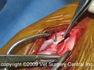
Surgery is the recommended treatment option for an avulsed long digital extensor tendon in the knee joint. An incision is made on the side of the knee over the affected tendon. If the avulsion fracture is fresh (occurred within 1 week), the bone and tendon may be reattached onto the femur bone with a screw. Because most avulsion fractures are chronic, the typical treatment involves suturing the long digital extensor tendon to the side of the joint capsule following removal of the bone fragment.

Results
Excellent results are achieved with sucessful surgery and full resolution of lameness may take 1 to 2 months. Uncommon complications may include swelling and infection. Some arthritis may develop in the knee joint, however this is usually not severe enough to cause significant clinical signs.
Aftercare
Following surgery, the patient will receive pain-relieving medication to ensure a comfortable recovery. A combination of nonsteroidal anti-inflammatory, local anesthetics and narcotics are used to control pain. Intravenous fluid therapy is administered to ensure that your companion will remain well hydrated after surgery. Most patients can go home on the day of surgery. At home, the incision should be checked for signs of infection. Your pet should not lick the incision, as this could open the incision or cause infection. If necessary, an Elizabethan collar can be placed on your companion to prevent licking and chewing at the surgical site. Exercise should be restricted for about 6 weeks after surgery. Rehabilitation therapy usually can be done at home; however, the surgeon may recommend that this is conducted by a professional rehabilitation therapist.
References
- Verhoeven G, VanRyssen B, Risselada M, van Bree H. Unusual presentation of an avulsion of the long digital extensor tendon in a dog: radiographic, computed tomographic, arthroscopic, surgical and histological findings. Vlaams Diergenesdundig Tijdschrift 76:438-42, 2007.
- Fitch RB, Wilson ER, Hathcock JT, Montgomery RD. Radiographic, computed tomographic and magnetic resonance imaging evaluation of a chronic long digital extensor tendon avulsion in a dog. Veterinary Radiology and Ultrasound 38, 177-181, 1997.
- Lammerding JJ, Noser GA, Brinker BO, Carrig CB. Avulsion fracture of the origin of the extensor digitorum longus muscle in three dogs. Journal of American Animal Hospital Association 12, 764, 1976.
- Olmstead ML, Butler HC. Surgical correction of avulsion of the long digital extensor in the dog. Veterinary Medicine/Small Animal Clinics 71, 608, 1976.
- Piermattei DL, Johnson KA. (2004). An Atlas of Surgical Approaches to the Bones and Joints of the Dog and the Cat. 4th ed. WB Saunders, Philadelphia, p 342-345. 6.Pond MJ. Avulsion of the extensor digitorum longus muscle in the dog. A report of four cases. Journal of Small Animal Practice 14, 785, 1973.
