

Key Points
Thymoma frequently has nonspecific clinical signs
Video Assisted Thoroscopic surgery is the best treatment option
Thymoma, if noninvasive carries a very good prognosis
What is the thymus?
The thymus is a an organ of the lymphatic system located within the chest cavity (just in front of the heart) that produces a subset of lymphocytes (white blood cells) called T-cells. These white blood cells are important to kill foreign cells, activate other immune cells to fight infection, and stop the immune response after a foreign cell or microbe has been eliminated. T-cells migrate away from the thymus to other parts of the body such as the lymph nodes, spleen, and the bloodstream. As the pet matures, the thymus has served its function, therefore it will wither away.
Tumors of the thymus gland
The thymus gland can develop a variety of cancers which most commonly include lymphoma and thymoma. Lymphoma is a cancer of the lymphocytes, whereas thymoma is a cancer that originates from the epithelial cells of the thymus. Thymoma can be either an invasive or noninvasive type of tumor. In fact, approximately 50% of the dogs have the non-invasive type of tumor which does not extend into the surrounding tissues and can be easily removed with surgery. The invasive thymoma is deeply rooted in surrounding structures such as the great vessels of the heart and the heart sac itself. Thymic carcinoma is a very rare, malignant tumor that spreads to other parts of the body.
Clinical signs
Thymoma most commonly affects medium to large breed dogs. Labradors and German shepherds are more commonly affected. Most pets with this condition are older, with the median age reported to be 11 years. Although likely coincidental, dogs having thymoma frequently have another type of cancer in their body. Only about 5% of the dogs with thymoma have hypercalcemia (high calcium level in the blood) due to the tumor. Hypercalcemia causes increased thirst, increased urination, decreased appetite, weakness, lethargy, and other nonspecific clinical signs. Most signs directly related to thymoma are nonspecific and may include decreased exercise tolerance, breathing difficulties, coughing, difficulty swallowing, and weight loss. The respiratory signs are due to enlargement of the tumor and compression of the windpipe within the chest cavity. Swallowing difficulties and regurgitation of fluid or food can be secondary to megaesophagus or compression of the esophagus within the chest. Paralysis of the voice box (laryngeal paralysis) can be secondary to nerve damage from the tumor. Invasive thymomas can cause cranial vena cava syndrome which manifests itself as swelling of the lower jaw, neck, forelimbs, and can result in fluid buildup in the chest (pleural effusion).
Para neoplastic syndromes secondary to thymoma include myasthenia gravis, hypercalcemia, aplastic anemia, immune-mediated diseases (immune mediated anemia, polymyositis dermatitis in cats), abnormal heartbeats due to inflammation of the heart muscle, and hypogammaglobulinemia.
Diagnosis
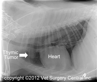
A presumptive diagnosis of thymoma is based on finding a mass on an x-ray in front of the heart. Other tumors in this area may also include lymphoma of the thymus, carcinoma of the thymus, branchial cyst, displaced thyroid or parathyroid tumors, aortic body tumors, tumors that have spread from elsewhere into this region, and sarcomas that extend from the chest wall. Definitive diagnosis requires a biopsy of the mass, which is commonly done after the tumor has been surgically removed. Fine needle biopsy prior to surgery can be performed and may be useful to diagnose tumors other than thymoma. One of the challenges, however, is that thymoma tumors also have lymphocytes in them, which may confuse the diagnosis with lymphoma. Other tests that are commonly performed in dogs that have a suspected thymoma include 3 view chest x-rays, advanced imaging such as CT scan or MRI, chest ultrasound, and bloodwork including a complete blood count and biochemistry profile, and an urinalysis.
Treatment
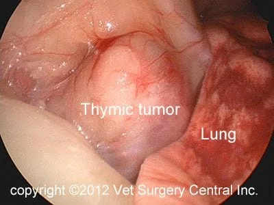
Surgery is the primary treatment for thymoma. Currently, in most surgical animal hospitals, tumors of the thymus gland are removed within an open surgical approach, which involves spreading the ribs or more commonly cutting the breast bone with a saw. I recommend to remove these tumors with video assisted thoroscopic surgery (VATS). During this procedure, a camera on a thin tube (thoroscope) is inserted into the chest via a small incision; this allows the surgeon to see the inside of the chest on a video monitor (see video right).
Two other small incisions are made to insert instruments into the chest cavity. With the use of a special cautery instrument called the Ligasure, the tumor is liberated from the surrounding tissues. The tumor is then placed in a surgical plastic bag and removed via one of the ports. A chest tube is placed after the surgery, regardless of whether it is done with the traditional open techniques or with VATS. The hospital stay following VATS is about 48 hours, versus 5 days with traditional surgery.
Radiation can be a very effective treatment for thymoma in dogs and cats. Approximately 75% of the patients will respond to this therapy. Radiation side effects may include inflammation of the lung and heart sac (pericardium). Radiation treatments are administered daily, 5 days a week, for a period of 18 to 21 treatments. As a result, radiation is only recommended in patients that have a tumor that cannot be surgically removed. Chemotherapy is largely ineffective for thymoma.
Prognosis
Most dogs that have a noninvasive thymoma removed are cured of their disease. In fact most of them died of unrelated causes. The overall one-year survival rate following thymectomy is 83%. If a patient has other complicating factors such as pneumonia or megaesophagus the prognosis may not be as good. The median survival time of dogs receiving radiation therapy is 248 days. Overall, the prognosis for dogs that have an invasive thymoma is guarded.
