

Key Points
Skin tumors can be benign or malignant
A biopsy of the tumor is needed to make the diagnosis prior to surgery
Surgical removal of the mass is the best option in most cases and the prognosis is dependant on the type of the tumor
Anatomy of the skin
The skin is made of 2 major layers, the epidermis and the dermis. Beneath the skin is a fatty layer with connective tissue called the subcutaneous tissue. The epidermis consists of epithelial cells (cells that are stuck together). The deep layer of the epidermis is actively growing, producing cells that gradually move to the surface of the skin. The dermis is a connective tissue layer that contains hair follicles, sebaceous gland (oil-producing glands) and sweat glands.
Definitions
Commonly, terms are used interchangeably and mean the same thing.
Mass is a lump or growth in the body’s tissues.
A tumor is a growth and can be benign or malignant
Neoplasm is a lump, mass, and tumor. This word means new (“neo”) growth (plasm) of cells.
Cancer is a tumor that by definition is malignant.
Malignant tumor has cells that are invasive into adjacent tissues and can metastasize (spread) to other parts of the body
Benign tumor means that it does not spread and it is just an annoying mass.
Metastatic tumor is a tumor that spread from another tumor in the body.
Metastasize means spreading of a tumor to another part of the body (typically through the blood stream or lymphatics)
Types of skin Masses that originate in the skin
Histiocytoma is a benign button tumor that typically is pink, raised is relatively hairless. Believed to potentially be induced by a virus, these tumors commonly will resolve spontaneously in most dogs. These can look identical to a mast cell tumor.
Mast cell tumor is considered a malignant tumor, that is the “imitator of tumors”. Therefore it can look like almost any type of tumor. In general, the “button” type of this tumor is a pink, raised, hairless tumor that may swell if it is pinched, rubbed or massaged.
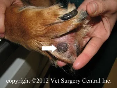
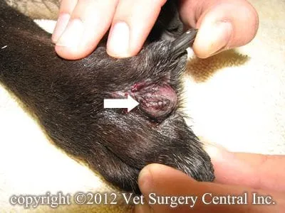
Basal cell carcinoma, more commonly found in cocker spaniels, is almost always a benign tumor of the skin and originates from the deep layer of the epidermis called the basal cell layer. Because this basal cell layer also surrounds hair follicles and sweat or sebaceous glands, they can also originate from these structures. This tumor located within skin of the head, neck and shoulders, is well-circumscribed, may be black in color.
Epidermal inclusion cyst is an accumulation of sebaceous gland secretions in a pocket within the skin or in the subcutaneous tissues. This is really not a tumor, but will feel like one. Sometimes these masses will rupture to the surface and the greasy central material of the mass will leak. These masses can become infected and be very painful to the pet.
Calcinosus circumscripta is a deposit of calcium within the skin. Secondary inflammation may also be seen. This mass, although not a cancer, may mimic the appearance of such. They commonly are very firm masses located almost anywhere on the body. These may be secondary to trauma, injections, and foreign bodies.
Melanomas, when located anywhere except in the mouth, lips, and toenail beds, have a low spread rate. When located in latter 3 sites, they commonly have a high tendency to spread to regional lymph nodes and the lungs. These tumors sometimes are black, yet other times do not have the black color as they have little pigment within the cells.
Plasmacytoma (extramedullary form) when found on the skin commonly is a benign mass. This type of tumor is malignant if it has spread from another part of the body. In order to prove that this type of tumor has not spread from the bone marrow to the skin, additional tests should be done to make sure that this the the case (bone marrow biopsy, serum electrophoresis, and evaluation of the the bones for tumor lesions with x-ray, CT, or bone scan). This tumor may appear to be a pink nodule within the skin.
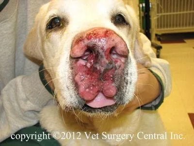
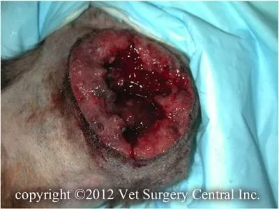
Squamous cell carcinoma s a malignant cancer of the skin. This tumor is firm, nodular, frequently ulcerated, and extend deeply into the dermis of the skin. This tumor is locally invasive, but the spread rate is relatively low to moderate, depending on its site on the body.
Hair follicle tumors exist in two main forms: the trichoepithelioma and the pilomatricoma. Both of these are benign. They are typically hairless and look like a basal cell carcinoma.
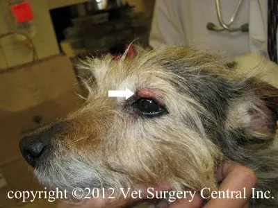
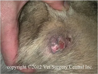
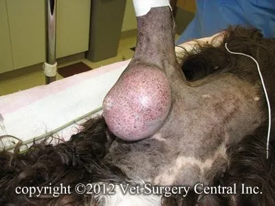
Sebaceous gland tumors originate from the oil producing cells in the skin. Sebaceous adenomas are benign, small, pink, lobulated (bumpy), and on the skin surface. Sebaceous epithelioma are benign masses that are most commonly found on the margin of the eyelids. Sebaceous gland carcinomas (see photo right) grow quickly, frequently are ulcerated, locally invasive and have a variable spread rate to other regions on the body.
Soft tissue sarcomas are malignant tumors that vary in their potential to metastasize. Grade 1 tumors have a 5 to 10% spread rate, grade 2 tumors have a 20 to 25% spread rate, and grade 3 tumors have a 50% spread rate. The growth rate may be slow or rapid and vary in their appearance. They commonly are not ulcerated, but as they outgrow their blood supply they can become ulcerated.
Diagnosis
Initial evaluation should first involve a fine needle biopsy of the tumor. A core biopsy of the tumor may collected if a fine needle biopsy is not providing a diagnosis. Chest x-rays and fine needle biopsy of regional lymph nodes is indicated in patients that have suspected cancer. Abdominal ultrasound may also be indicated to look for spread of the tumor to internal organs. Blood testing including a complete blood count and chemistry profile, and a urinalysis should be done to evaluate the patient’s internal organ health.
The day of surgery
Please make sure that your companion has been fasted prior to surgery and that the prescribed dose of Pepcid AC has been administered. The surgeon will contact you after the surgery to give you a progress report on your companion after the surgery. Our anesthesia and surgical team will prescribe a pain management program, both during and after surgery that will keep your companion comfortable. This will include a combination of general anesthesia, injectable analgesics, local anesthetics, oral analgesics and anti-inflammatory medication.
Treatment
The hair coat surrounding the tumor is clipped. Cancerous tumors mandate removing the tumor and about an inch or more of normal tissue surrounding the tumor. Benign tumors can be removed by only removing the tumor and a small amount of normal tissue surround the tumor.
Aftercare
Following surgery, the patient will receive pain-relieving medication to ensure a comfortable recovery. A combination of nonsteroidal anti-inflammatory, local anesthetics, and narcotics may be needed to control pain. At home, the incision should be checked for signs of infection. Your pet should not lick the incision, as this could open the incision or cause infection. If necessary, an Elizabethan collar can be placed on your companion to prevent licking and chewing at the surgical site. Antibiotics may be indicated after the surgery in some cases. Exercise should be restricted for about 3 weeks after surgery.
