

Key Points
Hypertrophic osteopathy is usually associated with a primary disease located within the abdomen or chest
Treatment of the primary problem will result in resolution of the bone thickening and pain
The prognosis is highly dependant on the inciting cause of hypertrophic osteopathy
Introduction
- Hypertrophic osteopathy is a condition that results in new bone formation (periosteal proliferation) on the bones of the limbs of an afflicted patient. Early in the course of HO, periosteal new bone production starts on the bones of the digits and paw bones and with time continues up the limb.
- Typically there is an underlying condition that is associated with this condition such as
- disease in the chest such as a rib tumor, primary lung tumor, metastatic lung tumor, rib tumor, lung abscess, parasite infection associated sarcoma of the esophagus, patent ductus arteriosus, congenital megaesophagus, infective endocarditis (infection of the heart valve), mycobacterial pneumonia, and bronchial foreign body with lobar pneumonia.
- disease in the abdomen such as kidney tumors, prostatic tumors and other masses within the abdomen
- The mechanism of formation of new bone on the limbs is not known, however, a disruption in the nerve signals results in a change in the blood supply of the covering of the bones (periosteum) that causes new bone to form on the surfaces of the bones.
- Resolution of the problem is typically seen once the underlying cause is removed (example: remove the primary tumor and the periosteal proliferation and lameness resolves)
Clinical signs
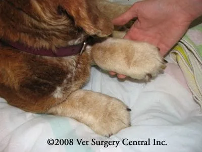
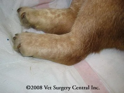
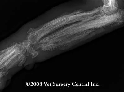
- Thickening of the limbs (see photos below)
- Lameness
- Pain upon palpation of the extremities
- Signs associated with the underlying problem such as coughing, weight loss, blood in the urine, loss of appetite
Diagnosis
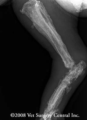
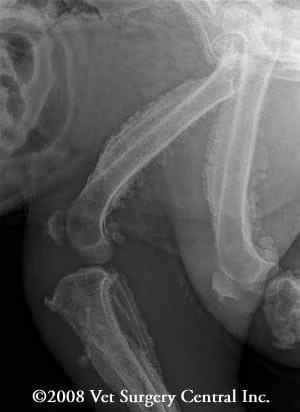
- The diagnosis of HO is made on finding the classic periosteal new bone production on the bones.
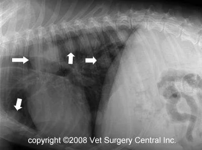
- A search for another tumor should always be done which includes chest radiographs, abdominal radiographs and ultrasound of the abdominal and visible structures in the chest.
- Your veterinarian may recommend a biopsy if a tumor is found in the chest or abdomen
- Blood work such as a CBC and chemistry, along with urine testing will also be recommended if your pet is going to have treatment of the condition (surgical removal of the primary tumor).
- The radiographs below demonstrate periosteal proliferation of the bones. The final radiograph, which is of the chest, demonstrates multiple lung tumors due to spread of tumor from the urinary bladder.
Treatment
- Treatment is only possible if the pet has an operable tumor
- By removing the tumor the periosteal proliferation will resolve and your pet will be relieved of the bone pain.
Prognosis
- If the primary tumor can be removed and has not spread, the prognosis is excellent, however if the tumor of primary inciting condition is not treatable, the prognosis is very poor.
References
- Mylonakis ME, Rallis T, Koutinas AF. Canine spirocercosis. Compend Contin Educ Vet. 2008 Feb;30(2):111-6.
- Grillo TP, Brandão CV, Mamprim MJ, de Jesus CM, Santos TC, Minto BW. Hypertrophic osteopathy associated with renal pelvis transitional cell carcinoma in a dog. Can Vet J. 2007 Jul;48(7):745-7.
- Chiang YC, Liu CH, Ho SY, Lin CT, Yeh LS. Hypertrophic osteopathy associated with disseminated metastases of renal cell carcinoma in the dog: a case report. J Vet Med Sci. 2007 Feb;69(2):209-12.
- Dunn ME, Blond L, Letard D, DiFruscia R. Hypertrophic osteopathy associated with infective endocarditis in an adult boxer dog. J Small Anim Pract. 2007 Feb;48(2):99-103.
- Foster WK, Armstrong JA. Hypertrophic osteopathy associated with pulmonary Eikenella corrodens infection in a dog. J Am Vet Med Assoc. 2006 May 1;228(9):1366-9.
- Anderson TP, Walker MC, Goring RL. Cardiogenic hypertrophic osteopathy in a dog with a right-to-left shunting patent ductus arteriosus. J Am Vet Med Assoc. 2004 May 1;224(9):1464-6, 1453.
- Watrous BJ, Blumenfeld B. Congenital megaesophagus with hypertrophic osteopathy in a 6-year-old dog. Vet Radiol Ultrasound. 2002 Nov-Dec;43(6):545-9.
- Harrus S, Waner T, Aizenberg, Safra N, Mosenco A, Radoshitsky M, Bark H. Development of hypertrophic osteodystrophy and antibody response in a litter of vaccinated Weimaraner puppies. J Small Anim Pract. 2002 Jan;43(1):27-31.
- Peeters D, Clercx C, Thiry A, Hamaide A, Snaps F, Henroteaux M, Ogilvie GK, Day MJ. Resolution of paraneoplastic leukocytosis and hypertrophic osteopathy after resection of a renal transitional cell carcinoma producing granulocyte-macrophage colony-stimulating factor in a young Bull Terrier. J Vet Intern Med. 2001 Jul-Aug;15(4):407-11.
- Barrand KR, Scudamore CL. Canine hypertrophic osteoarthropathy associated with a malignant Sertoli cell tumour. J Small Anim Pract. 2001 Mar;42(3):143-5.
- Miller C. Hypertrophic osteodystrophy in a Great Dane puppy. Can Vet J. 2001 Jan;42(1):63-6.
- Wylie KB, Lewis DD, Pechman RD, Cho DY, Roy A. Hypertrophic osteopathy associated with Mycobacterium fortuitum pneumonia in a dog. J Am Vet Med Assoc. 1993 Jun 15;202(12):1986-8.
- Hesselink JW, van den Tweel JG. Hypertrophic osteopathy in a dog with a chronic lung abscess. J Am Vet Med Assoc. 1990 Mar 1;196(5):760-2.
- Caywood DD, Kramek BA, Feeney DA, Johnston GR. Hypertrophic osteopathy associated with a bronchial foreign body and lobar pneumonia in a dog. J Am Vet Med Assoc. 1985 Apr 1;186(7):698-700.
